Xiaoxu Yang1, Qi Yang2, Jiangang Duan2, Zhaoyang Fan3, Fang Wu4, and Xunming Ji4
1Chaoyang Hospital, Beijing, China, 2Chaoyang Hospitao, Beijing, China, 3Cedars Sinai Medical Center, Los Angeles, CA, United States, 4Xuanwu Hospital, Beijing, China
1Chaoyang Hospital, Beijing, China, 2Chaoyang Hospitao, Beijing, China, 3Cedars Sinai Medical Center, Los Angeles, CA, United States, 4Xuanwu Hospital, Beijing, China
BTI provided high diagnostic value of BTI and another advantage of accurate staging (acute, subacute and chronic), achieving more thrombosed segments of definite stage, which is important for clinical strategy.
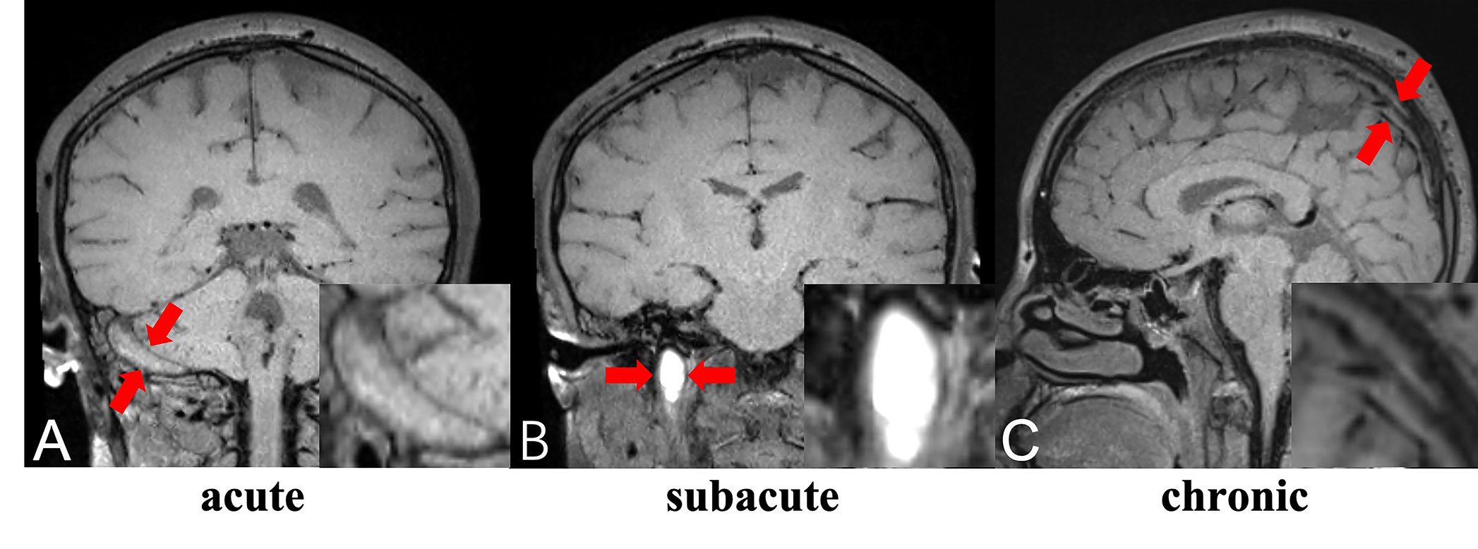
BTI images of a 24-year-old woman with multiple thrombosed segments of varied stage. Coronary BTI image showed isointense signal indicative of acute thrombus in right sigmoid sinus (red arrows in A). Coronary BTI image showed hyperintense signal indicative of sub-acute thrombus in right jugular vein (red arrows in B). Sagittal BTI image showed isointense signal with flow voids indicative of chronic thrombus (red arrows in C) in right sigmoid sinus.
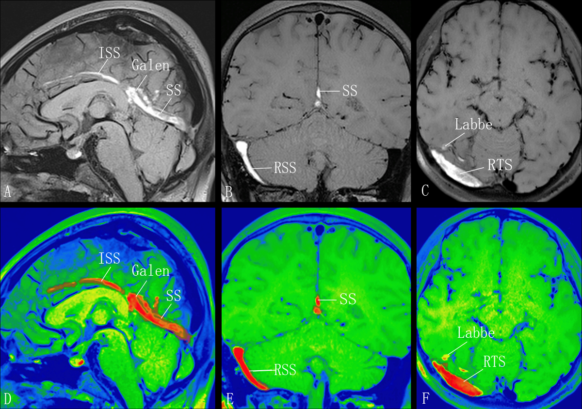
BTI images of a 13-year-old female with suspected CSVT. Sagittal, coronal, axial images of BTI (A, B, and C) and corresponding color graphs made by commercial software (Object Research System, Montreal, Quebec, Canada) depicted the hyperintense signal of subacute thrombi in Inferior sagittal sinus, vein of Galen, straight sinus, right sigmoid sinus and right transverse sinus.