Tongtong Sun1, Yu Zhang1, Tongtong Li1, Yuan Luo2, Jinxia Zhu3, Cheng Cheng4, and Hongyan Ni5
1Radiology, First Central Clinical College, Tianjin Medical University, Tianjin, China, 2Radiology, China-Japan Friendship Hospital, Beijing, China, 3MR Collaboration, Siemens Healthcare, Beijing, China, 4MR Application, Siemens Healthcare, Beijing, China, 5Radiology, Tianjin First Central Hospital, Tianjin, China
1Radiology, First Central Clinical College, Tianjin Medical University, Tianjin, China, 2Radiology, China-Japan Friendship Hospital, Beijing, China, 3MR Collaboration, Siemens Healthcare, Beijing, China, 4MR Application, Siemens Healthcare, Beijing, China, 5Radiology, Tianjin First Central Hospital, Tianjin, China
PETRA-MRA significantly improved the uniformity and CR of the cavernous segment of ICA, although it is inferior in both SNR and CNR. This suggests that PETRA-MRA could provide a reasonable alternative to reduce the loss of signal from curved vessels.

Fig.2
MIP
images of a 32 years old healthy man (a, b). TOF-MRA reveals signal loss in the
cavernous segment of the internal carotid artery, but the signal
is uniform in the same position for PETRA-MRA(box). MIP images of a 41 years old healthy
woman (c, d). In addition to the loss of signal, TOF-MRA also shows a coloboma
in the vascular profile, but in PETRA-MRA, it is smooth and complete (arrow).
Source images of a 55 years old healthy woman (e, f). Low signal intensity
occurs in the center of the lumen and part of the boundary in TOF-MRA, but it
is not visualized for PETRA-MRA (double-headed arrow).
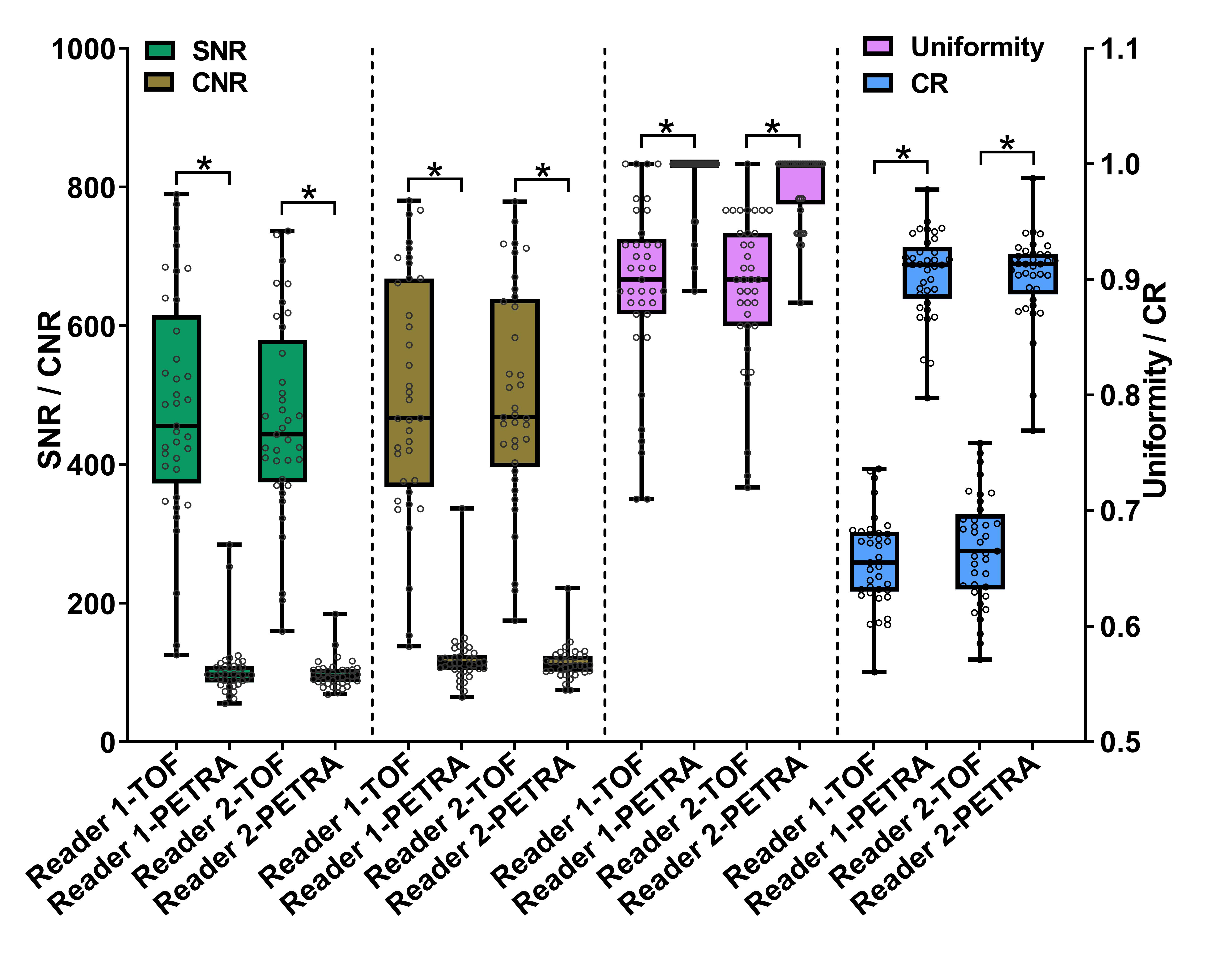
Fig.1 All
points, median, 25% quartile, 75% quartile, the minimum, and the maximum value
of 37 subjects on SNR, CNR, uniformity, as measured by Reader 1 and Reader 2,
are shown in the box-and-whisker plot. The plot reveals higher SNR and CNR,
lower CR and uniformity in TOF-MRA compared with PETRA-MRA. The differences
between the two MRA images in these four indicators are statistically
significant (all P < 0.001, which is indicated by * in the plot).