Cheng Zhong1, Niranjan Balu2, Chun Yuan2, and Zhensen Chen2,3
1Fudan University, Shanghai, China, 2Department of Radiology, University of Washington, Seattle, WA, United States, 3Center for Biomedical Imaging Research, School of Medicine, Tsinghua University, Beijing, China
1Fudan University, Shanghai, China, 2Department of Radiology, University of Washington, Seattle, WA, United States, 3Center for Biomedical Imaging Research, School of Medicine, Tsinghua University, Beijing, China
Purpose a segmentation method based on dMRA,
which uses the spatial-temporal correlation between vessel voxels to improve vessel
segmentation.
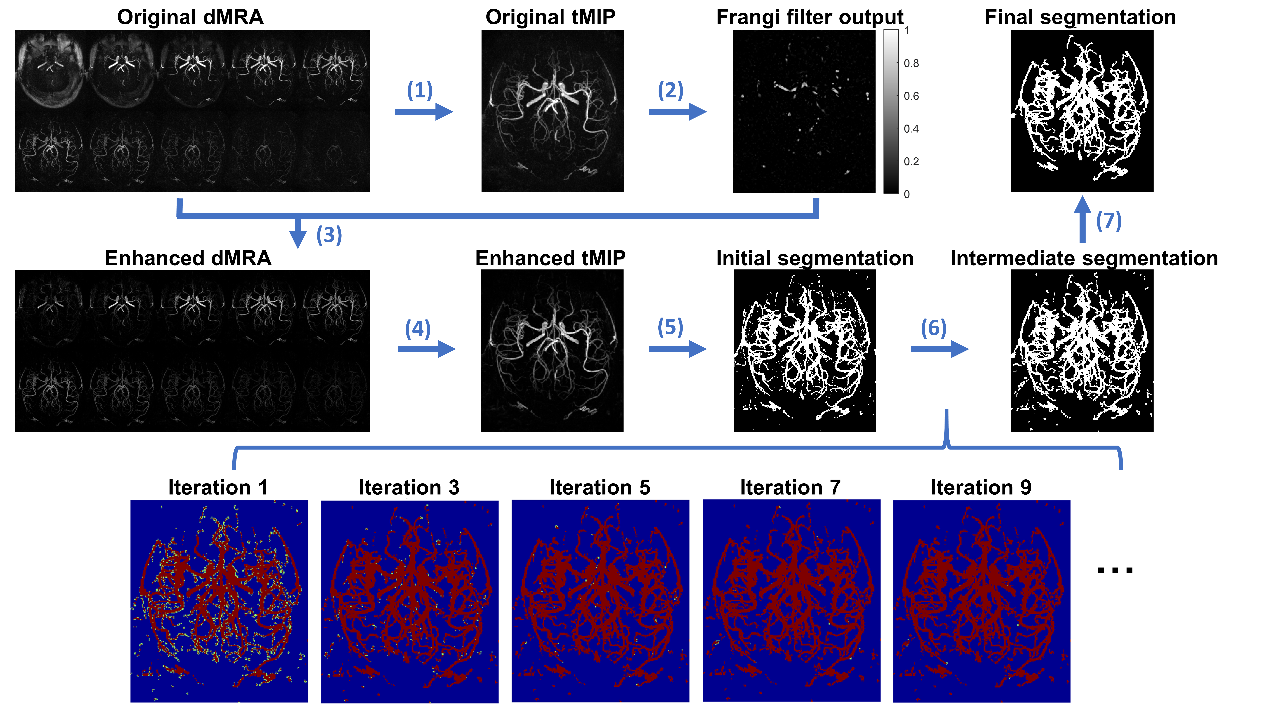
Figure 2. Pipeline of the proposed segmentation method (1) Perform
maximum intensity projection along the temporal dimension of dMRA to generate tMIP image volume. (2) Enhance columnar structure with Frangi filter. (3) Use Frangi filter output to
enhance dMRA. (4) Regenerate tMIP from the enhanced dMRA. (5) Use threshold method to yield initial
segmentation. (6) Iteratively expand the
segmentation based on spatial-temporal correlation between voxels. Light color
in the bottom images indicates new vessel pixels found by the iteration. (7) Cleanup small connected components.
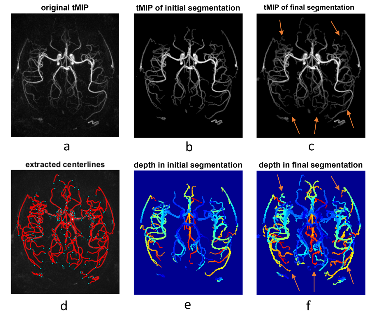
Figure 3. Vascular segmentation results by the proposed method on volunteer #1 (female, 24 years). The images in the upper row are MIP of tMIP along feet-head direction. To better distinguish the vessels, the depth images calculated from MIP of the final segmentations, instead of the binary MIP, are shown in (e) and (f). (d) shows the extracted and smoothed centerline obtained from the final segmentation. Example distal vessels extracted by correlation-based method but not by the threshold method are indicated with arrows.