Matthijs de Buck 1, Peter Jezzard1, and Aaron Hess2
1Wellcome Centre for Integrative Neuroimaging, FMRIB Division, Nuffield Department of Clinical Neurosciences, University of Oxford, Oxford, United Kingdom, 2Oxford Centre for Clinical Magnetic Resonance Research, Department of Cardiovascular Medicine, University of Oxford, Oxford, United Kingdom
1Wellcome Centre for Integrative Neuroimaging, FMRIB Division, Nuffield Department of Clinical Neurosciences, University of Oxford, Oxford, United Kingdom, 2Oxford Centre for Clinical Magnetic Resonance Research, Department of Cardiovascular Medicine, University of Oxford, Oxford, United Kingdom
Using small calibration regions of 12x12
lines with a polynomial order of about 2 is optimal for 3D TOF-MRA at 7T. This
is consistent for different acceleration factors, although optimization is
increasingly important at higher accelerations.
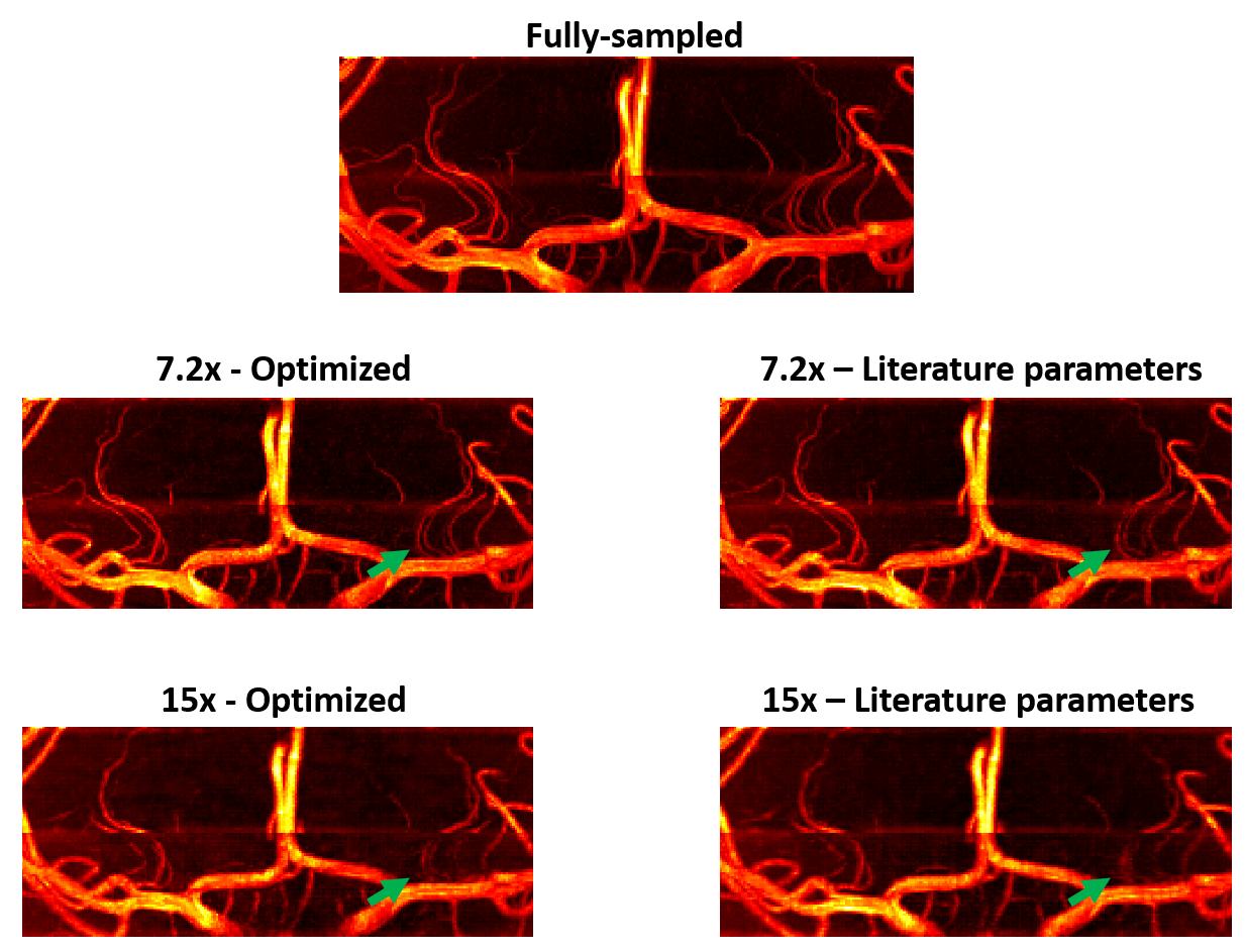
Figure 5: Coronal
MIPs of the sub-volume containing the lenticulostriate arteries in the same
reconstructions as in Figure 4. Green arrows indicate examples of improved
visibility and sharpness of the arteries when using optimized sampling
parameters, for both acceleration factors.
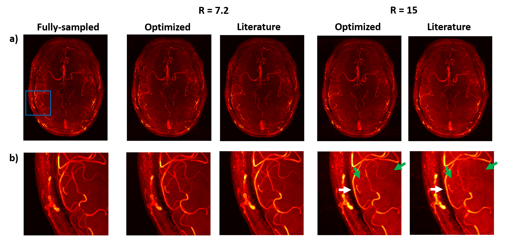
Figure 4: Axial MIPs of reconstructions
from fully-sampled and prospectively undersampled datasets. Undersampled datasets are shown
for R=7.2 and R=15 with optimized (calib = 12, pp = 2.0) and literature-based
(calib = 32, pp = 2.4) undersampling parameters. (a) Axial MIPs of the
entire imaging volume. (b) Close-ups of the MIPs (blue box in fig. (a))
for improved visibility of smaller vessels. Especially for R=15, the optimized
acquisition shows improved visibility (green arrows) and sharpness (white
arrows) of the smaller vessels.