Maoxue Wang1, Yi Wang1, Yongbo Yang1, Yongbo Yang1, Ming Li1, Jilei Zhang1, and Bing Zhang1,2
1The Affiliated Drum Tower Hospital of Nanjing University Medical School, Nanjing, China, 2Institute of Brain Science, Nanjing University Nanjing, Nanjing, China
1The Affiliated Drum Tower Hospital of Nanjing University Medical School, Nanjing, China, 2Institute of Brain Science, Nanjing University Nanjing, Nanjing, China
The
4D-sPACK provided better performance than 3D TOF MRA in the treatment
evaluation of patients after bypass surgery, and it had high consistency with
DSA.
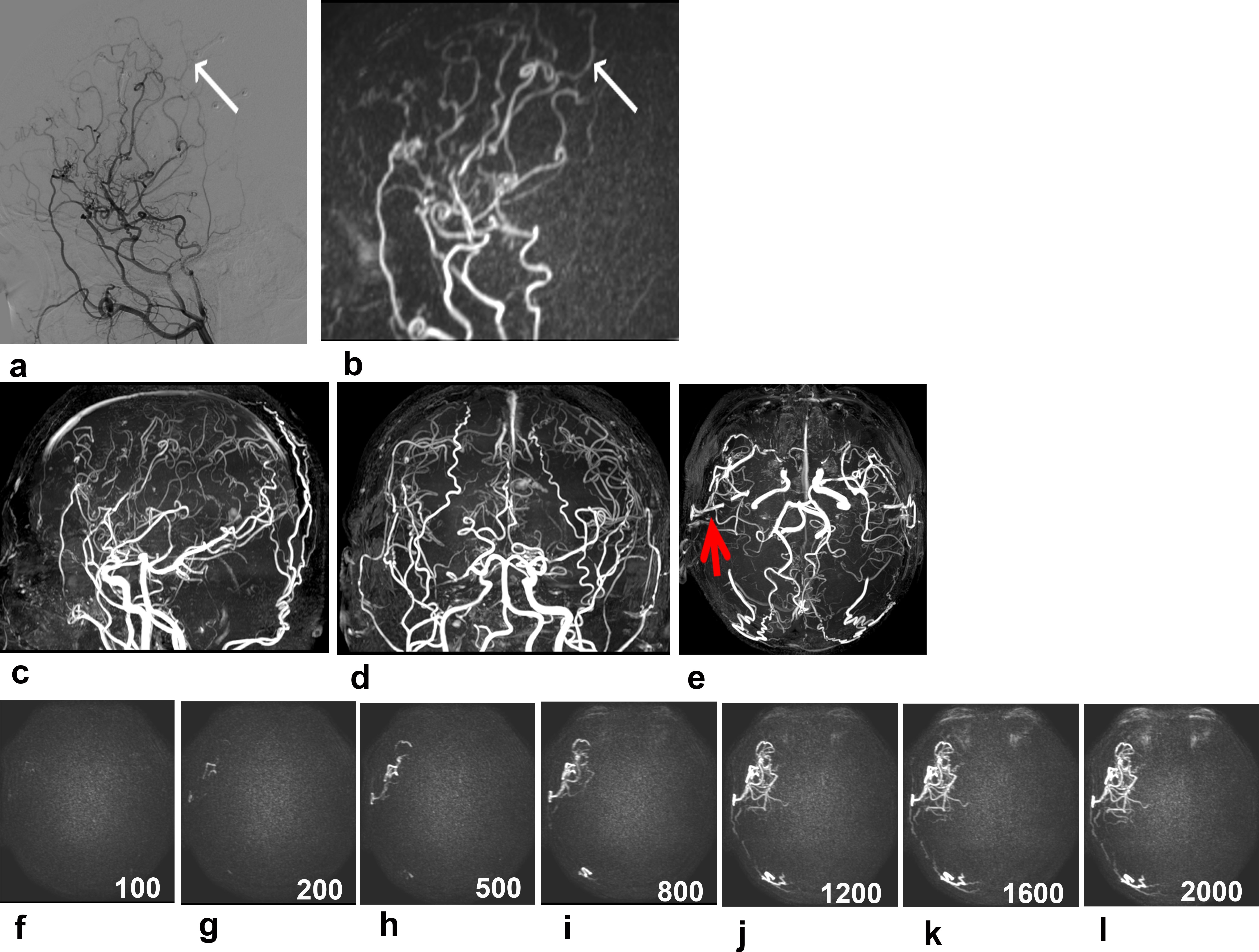
Figure
1. A 33-year-old male patient with bilateral MMV after
bilateral bypass surgery. The vasculopathy was observed approximately 35 months
after the surgery was performed on the right side. Intracranial collaterals originating
from the right external carotid artery were clearly shown on sagittal 4D-sPACK
(b), consistent with DSA (a); however, they were not clearly observed on axial,
coronal, and sagittal images (c–e) obtained on TOF MRA because of overlap with other vessels.
The anastomosis was shown on axial TOF MRA image (e, red arrow) and axial 4D-sPACK (f–i).
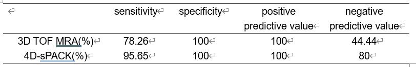
Table 1.
Results for anastomosis patency in 4D-sPACK and 3D TOF MRA images using DSA
as the gold standard.