Haruyuki Fukuchi1,2,3, Toshiya Akatsu4, Kei Fukuzawa3, Nao Takano4, Yutaka Ikenouchi2, Michimasa Suzuki2, Kohji Kamagata2, Akihiko Wada2, Osamu Abe1, and Shigeki Aoki2
1Radiology, Department of Radiology, Graduate School of Medicine, University of Tokyo, Tokyo, Japan, 2Department of Radiology, Juntendo University Graduate School of Medicine, Tokyo, Japan, 3Department of Radiology, Toranomon Hospital, Tokyo, Japan, 4Department of Radiology, Juntendo University Hospital, Tokyo, Japan
1Radiology, Department of Radiology, Graduate School of Medicine, University of Tokyo, Tokyo, Japan, 2Department of Radiology, Juntendo University Graduate School of Medicine, Tokyo, Japan, 3Department of Radiology, Toranomon Hospital, Tokyo, Japan, 4Department of Radiology, Juntendo University Hospital, Tokyo, Japan
The novel method, Variable TI improves non-contrast ASL based UTE
4D-MRA. This
method offered a higher signal intensity up to 50 % without reducing structural
information and improved visualization of arteries in late phases.
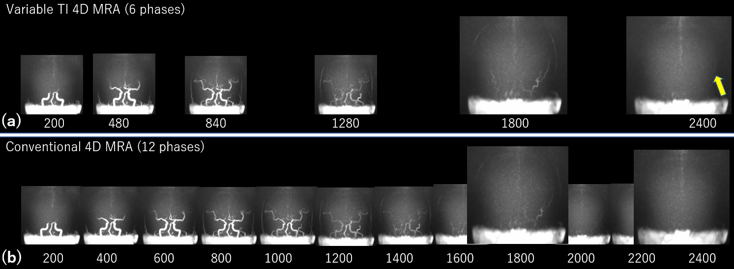
Figure .5. Comparison between the conventional UTE 4D MRA
and VTI UTE 4D MRA. Note that different time scales were used to display the
full range of information in each dataset and the times shown are relative to
start of labeling. (a) VTI UTE 4D MRA with 6 phases. (b) Conventional UTE 4D
MRA with 12 phases. The sequence chart is as shown in Figure 3. VTI UTE 4D MRA
better image quality comparing to conventional method without reducing
structural information. The late phase of image with VTI method has better visibility of arteries compared to conventional method.
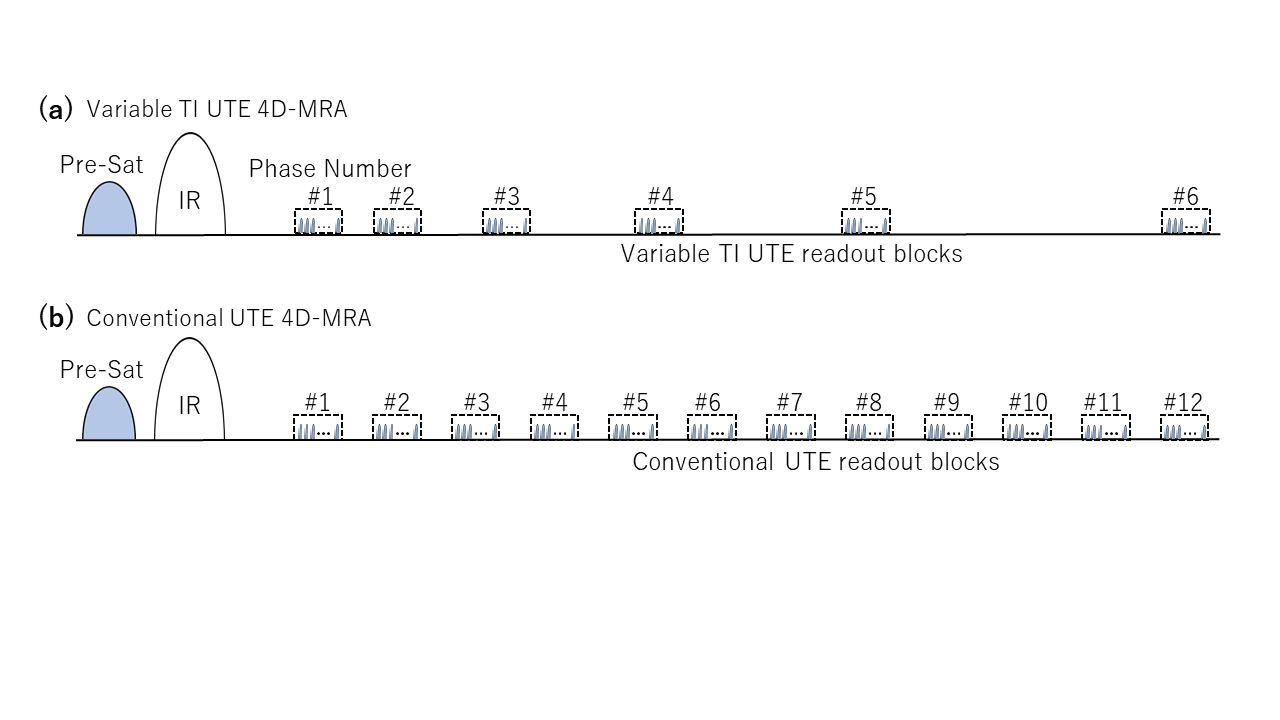
Figure .3. The graphical sketches showing UTE 4D-MRA pulse sequences used in the phantom
and volunteer study. (a) Variable TI UTE 4D-MRA, where the TI intervals were
increased with a manner of progression of difference, TI = 200, 480, 840, 1280,
1800, and 2400 ms, (b) conventional UTE 4D-MRA, 12 readout blocks from 200 to 2400
ms each blocks having 200 ms of delay time from the previous blocks. Each
readout block consists of 53 series of UTE acquisitions. The data from each
readout blocks were used to construct a single image.