Do-Wan Lee1, Hwon Heo2, Jae-Im Kwon3, Yeon Ji Chae2, Joongkee Min3, Monica Young Choi2, Chul‐Woong Woo3, Dong‐Cheol Woo2,3, Kyung Won Kim1, Jeong Kon Kim1, Hyo Jeong Chin4, and Dong‐Hoon Lee4
1Department of Radiology, Asan Medical Center, University of Ulsan College of Medicine, Seoul, Korea, Republic of, 2Department of Convergence Medicine, Asan Medical Center, University of Ulsan College of Medicine, Seoul, Korea, Republic of, 3Convergence Medicine Research Center, Asan Institute for Life Sciences, Asan Medical Center, Seoul, Korea, Republic of, 4Department of Radiological Science, College of Health Sciences, Yonsei University, Wonju, Korea, Republic of
1Department of Radiology, Asan Medical Center, University of Ulsan College of Medicine, Seoul, Korea, Republic of, 2Department of Convergence Medicine, Asan Medical Center, University of Ulsan College of Medicine, Seoul, Korea, Republic of, 3Convergence Medicine Research Center, Asan Institute for Life Sciences, Asan Medical Center, Seoul, Korea, Republic of, 4Department of Radiological Science, College of Health Sciences, Yonsei University, Wonju, Korea, Republic of
This study aimed to
visualize and quantitatively evaluate hippocampal glutamate changes in a rat
model of depression using in vivo proton magnetic resonance spectroscopy
(1H-MRS) and glutamate chemical exchange saturation transfer imaging
(GluCEST).
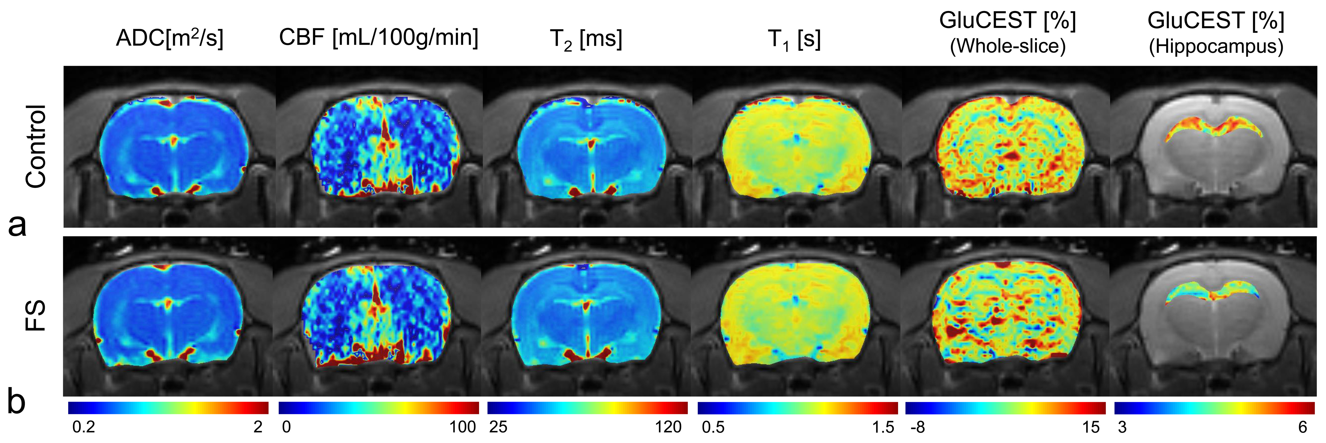
Figure 4. Reconstructed
typical multi-parametric MR images and GluCEST maps of the control (a) and FS
(b) rats. ADC, apparent diffusion coefficient; CBF, cerebral blood flow; FS,
forced swimming test group; GluCEST, glutamate-weighted chemical exchange
saturation transfer
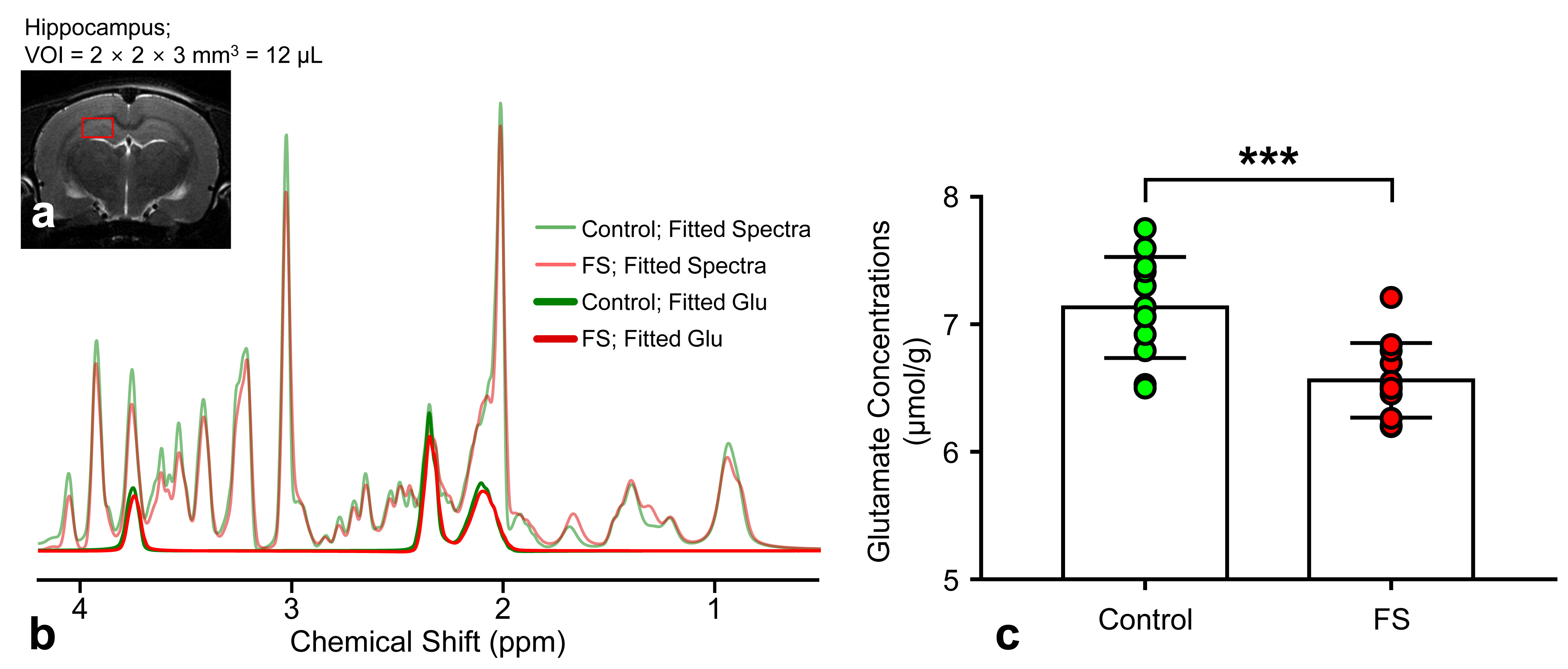
Figure 2. LCModel
fitted results of the 1H-MRS spectra in the hippocampal region
acquired from representative FS and control rats (a and b). The bar chart with
data points shows the mean glutamate concentration, and vertical lines on each
of the bars represent the standard deviation of the mean values (FS, forced
swimming test group; ppm, part per million; VOI, volume of interest; green
color: control group; red color: FS group; significance levels: ***p < 0.001).