Faye McKenna1, Yu Veronica Sui2, Hillary Bertisch2, Donald Goff2, and Mariana Lazar2
1Radiology, New York University School of Medicine, New York, NY, United States, 2New York University School of Medicine, New York, NY, United States
1Radiology, New York University School of Medicine, New York, NY, United States, 2New York University School of Medicine, New York, NY, United States
We employed NODDI-B, DKI and classical DTI and found that PSD and PSD sub-types had significantly different diffusivity especially in the secondary direction in sub-cortical WM ROIs compared to healthy controls which related to episodic and working memory and several schizotypal traits.
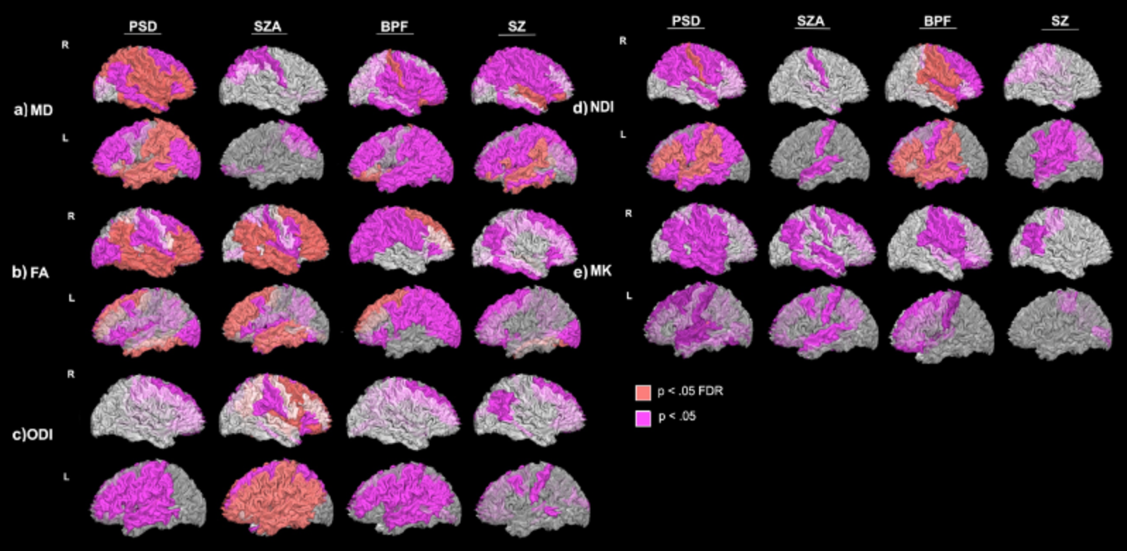
Figure 2. Group differences in non-directional dMRI metrics: a) MD, b) FA, c) ODI, d) NDI, and e) MK in the sub-cortical white matter. Results are presented by projecting WM areas of difference onto the corresponding sub-cortical WM surface. T-tests were conducted between HC and PSD and PSD subtypes: schizoaffective (SZA), schizophrenia (SZ) and bipolar with psychotic features (BPF). Group differences that reached significance at the FDR BH q < .05 correction-level are shown in pink, while group differences that reached significance at p < .05 trend-level are shown in purple.
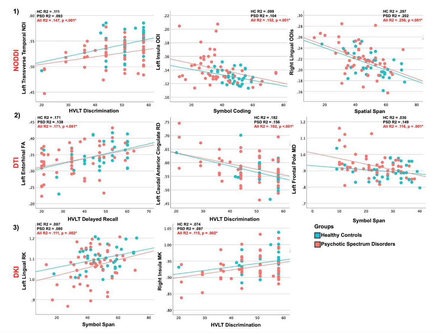
Figure 3. Representative significant Pearson’s correlations between dMRI metrics: 1) NODDI NDI, ODI and ODIs, 2) DTI FA, RD and MD, and 3) DKI RK and MK and several tests of verbal learning and memory (HVLT), processing speed (symbol coding) and working memory (spatial span, symbol span) in both PSD and HC groups. Tests that reached FDR BH q < .05 correction-level are noted with an asterisk (*).