Ting Su1, Jia ying Gong2, Shao juan Qiu1, Pan Chen1, Guan mao Chen1, Jun jing Wang3, Li Huang1, and Ying Wang1
1Medical Imaging Center, First Affiliated Hospital of Jinan University, Guangzhou, China, 2Department of Radiology, Six Affiliated Hospital of Sun Yat-sen University, Guangzhou, China, 3Department of Applied Psychology, Guangdong University of Foreign Studies, Guangzhou, China
1Medical Imaging Center, First Affiliated Hospital of Jinan University, Guangzhou, China, 2Department of Radiology, Six Affiliated Hospital of Sun Yat-sen University, Guangzhou, China, 3Department of Applied Psychology, Guangdong University of Foreign Studies, Guangzhou, China
AN
patients displayed decreased and increased functional activity in the ACC, MCC,
and parahippocampal gyrus, decreased GMV in the MCC and left inferior parietal. This
multimodal meta-analysis identified functional activity and gray matter
reductions in the ACC and MCC in patients with AN.
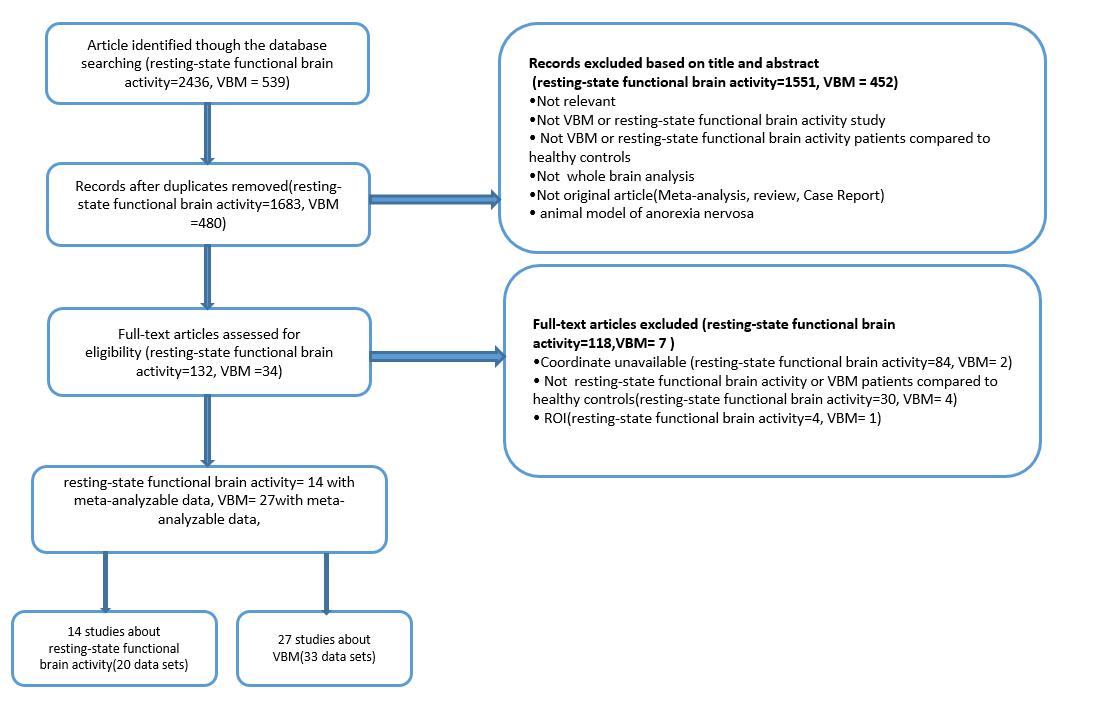
Fig. 1 Flow chart of Meta-analysis of
resting-state functional imaging and VBM studies of patients with AN
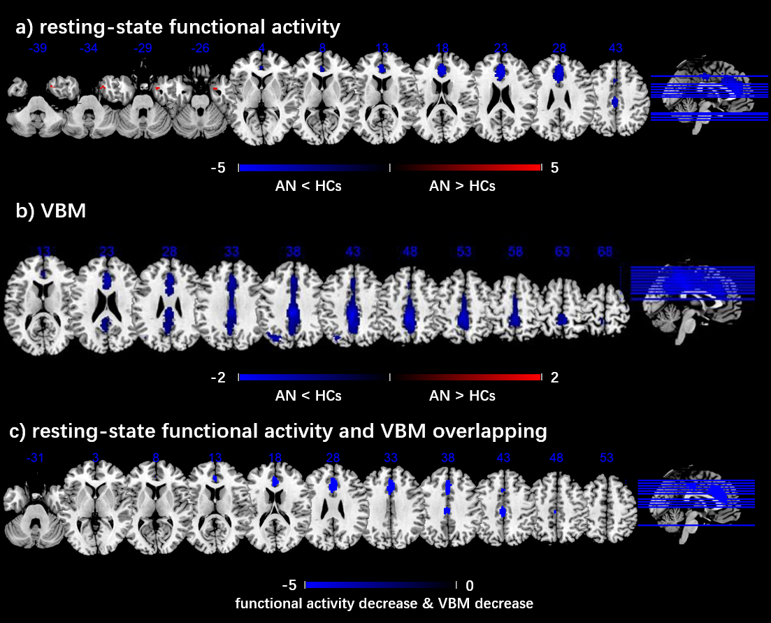
Fig. 2 Meta-analyses results regarding a) resting-state functional activity
difference between AN and HCs, b) GMV difference between
AN and HCs, c) conjunction of resting-state
functional activity differences and GMV differences. Areas with
decreased resting-state functional activity value or GMV value are displayed in
blue, and areas with increased resting-state functional activity value or GMV
value are displayed in red. The color bar indicates the maximum and minimum
SDM-Z values.