Koen P.A. Baas1, Simon Körver2, Bram F. Coolen3, Gustav J. Strijkers3, Carla E.M. Hollak2, and Aart J. Nederveen1
1Radiology and Nuclear Medicine, Amsterdam UMC, Amsterdam, Netherlands, 2Endocrinology and Metabolism, Amsterdam UMC, Amsterdam, Netherlands, 3Biomedical Engineering and Physics, Amsterdam UMC, Amsterdam, Netherlands
1Radiology and Nuclear Medicine, Amsterdam UMC, Amsterdam, Netherlands, 2Endocrinology and Metabolism, Amsterdam UMC, Amsterdam, Netherlands, 3Biomedical Engineering and Physics, Amsterdam UMC, Amsterdam, Netherlands
ADC
values in Fabry patients were significantly higher within white matter lesions
compared to healthy white matter, and increased further with lesion age.
Moreover, before lesions were detected on FLAIR images, we already found abnormal
ADC values in these specific regions.
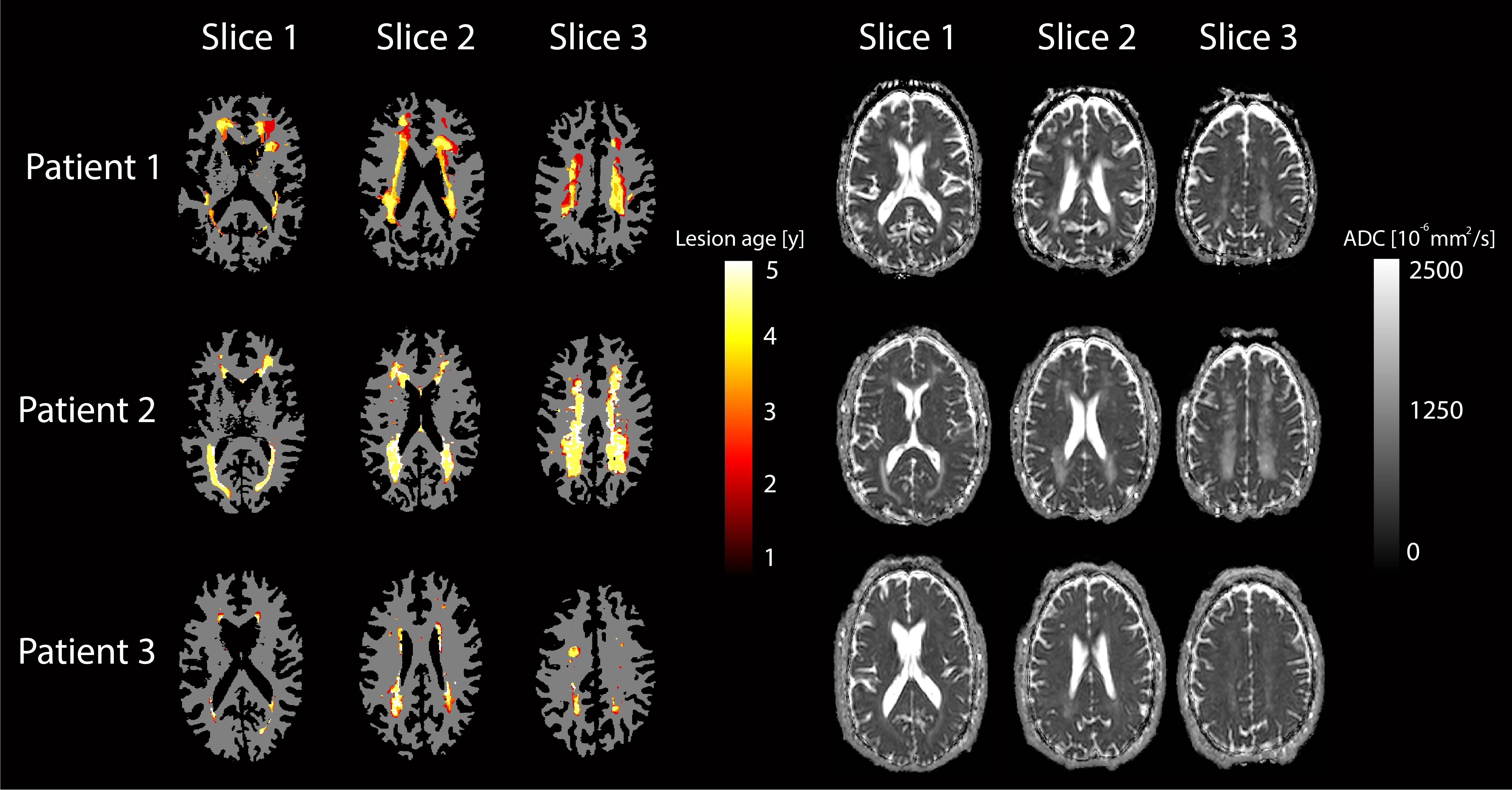
Figure 1:
lesion progression in three patients projected on WM segmentations (left) and
corresponding slices of ADC maps (right). Colors indicate the age of the
lesion. Only four time points were available for patient one and does therefore
not show lesion age of five years. Patient two and three had five available
time points.
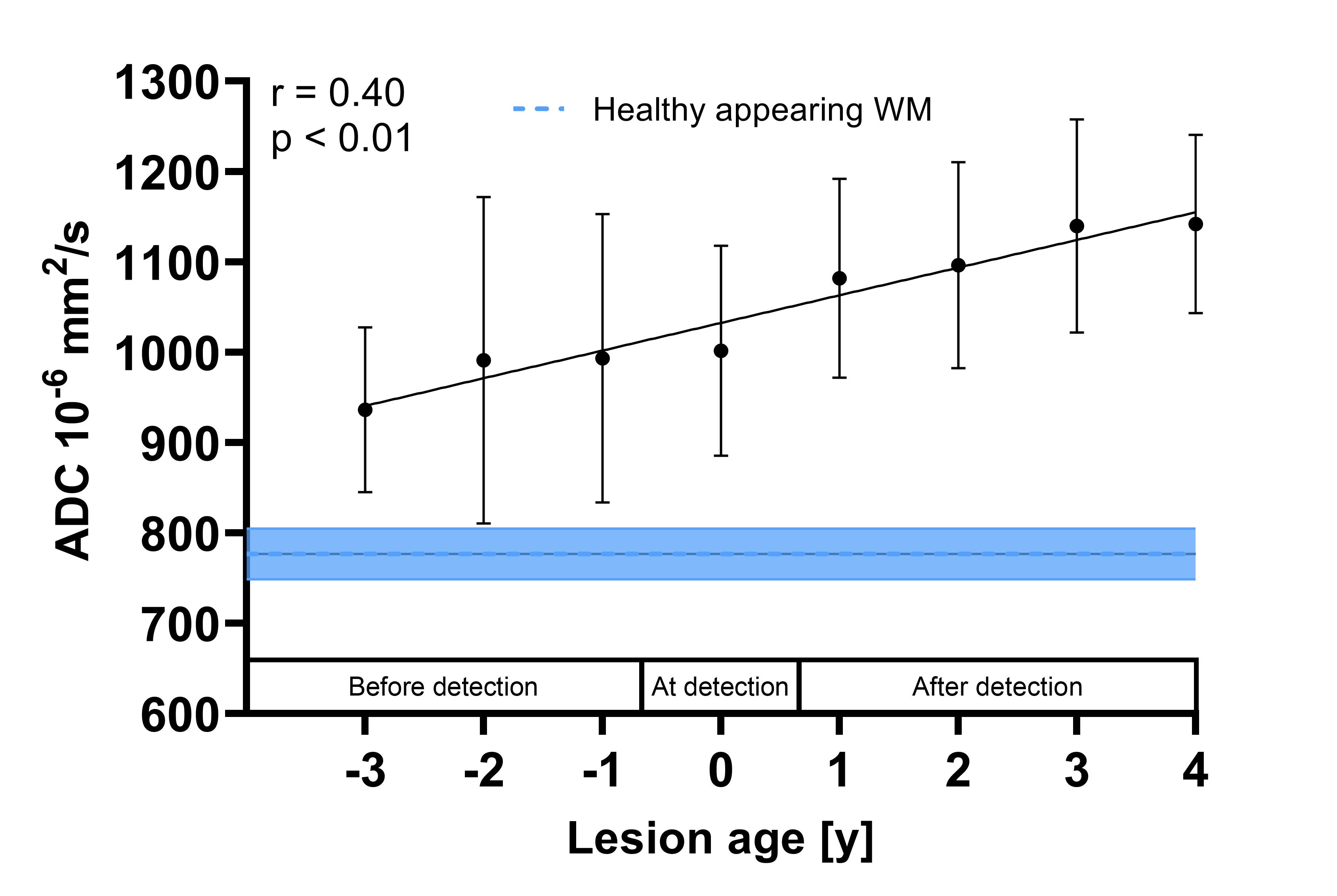
Figure 2: averaged
ADC values within newly detected WML as a function of lesion age. WMLs that
were present at the first time point were not included because the age of these
lesions cannot be determined. Error bars denote standard deviations. Linear
regression of all individual newly detected lesion areas after each year showed
a significant correlation between lesion age and ADC.