Teddy Salan1, Varan Govind1, and Sameer Vyas2
1University of Miami, Miami, FL, United States, 2Post Graduate Institute of Medical Education & Research, Chandigarh, India
1University of Miami, Miami, FL, United States, 2Post Graduate Institute of Medical Education & Research, Chandigarh, India
We applied free-water eliminated
diffusion tensor imaging (FWE-DTI) to calculate the
free-water volume content fraction (fFW)
in the brains of HE and healthy control subjects. We found significant fFW
increases among HE patients indicating low-grade edema and glial swelling.
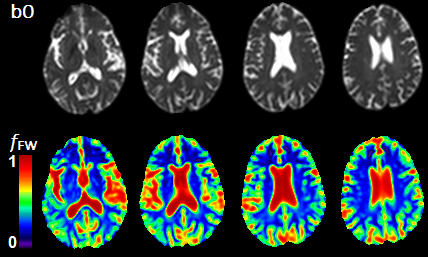
Axial slices from the brain of an HE subject showing b0 images (top row) and the corresponding the fFW maps (bottom row).
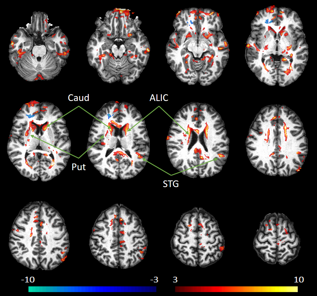
Voxelwise z-score maps comparing fFW values from the brain of an HE subjects with the mean values of the control group. The z-scores are overlaid on a template T1 image in MNI space.