Manuel Blesa Cábez1, Thijs Dhollander2, Victoria J Monnelly1, Alan J Quigley3, Scott I Semple4, Mark E Bastin4, and James P Boardman1
1MRC Centre for Reproductive Health, University of Edinburgh, Edinburgh, United Kingdom, 2Developmental Imaging, Murdoch Children's Research Institute, Melbourne, Australia, 3Department of Radiology, Royal Hospital for Sick Children, Edinburgh, United Kingdom, 4Edinburgh Imaging, University of Edinburgh, Edinburgh, United Kingdom
1MRC Centre for Reproductive Health, University of Edinburgh, Edinburgh, United Kingdom, 2Developmental Imaging, Murdoch Children's Research Institute, Melbourne, Australia, 3Department of Radiology, Royal Hospital for Sick Children, Edinburgh, United Kingdom, 4Edinburgh Imaging, University of Edinburgh, Edinburgh, United Kingdom
Prenatal exposure to methadone is associated with microstructural alterations in major white matter tracts, assessed by a reduction in fiber-bundle cross-section and fiber density and cross-section apparent soon after birth.
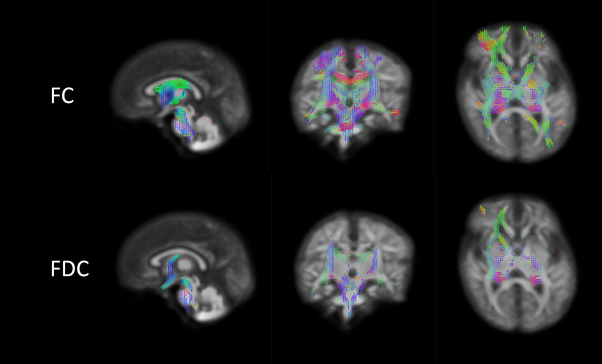
Figure 1: The top panels show
the areas where the prenatal methadone exposed group has lower FC and the
bottom panels show and the areas where they have lower FDC values. All the
results are overlaid to the WM FOD template for reference. Axial and coronal views
follow radiological convention.
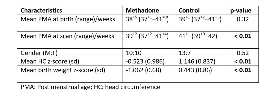
Table 1: Infant characteristics.