Ryckie George Wade1, Timothy T Griffiths1, Robert Flather1, Irvin Teh1, Hamied A Haroon2, David Shelley3, Sven Plein1, and Grainne Bourke3
1University of Leeds, Leeds, United Kingdom, 2University of Manchester, Manchester, United Kingdom, 3Leeds Teaching Hospitals, Leeds, United Kingdom
1University of Leeds, Leeds, United Kingdom, 2University of Manchester, Manchester, United Kingdom, 3Leeds Teaching Hospitals, Leeds, United Kingdom
This proof-of-concept study shows that, throughout the length of the ulnar nerve, fractional anisotropy and radial diffusivity are significantly different between asymptomatic adults and adults with cubital tunnel syndrome.
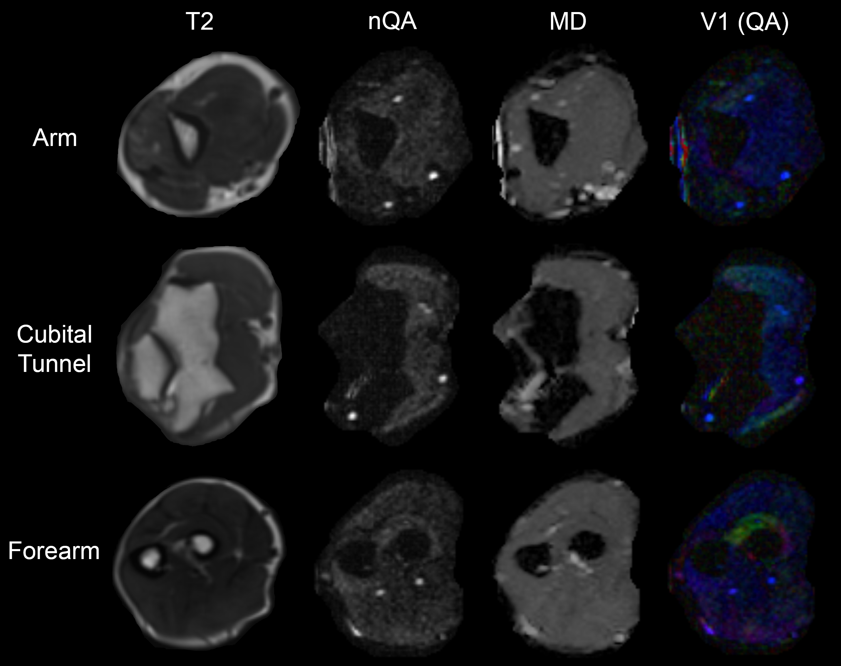
Figure 1. Data derived from a healthy control. The rows show data from the arm, cubital tunnel and forearm. The columns contain T2-weighted scans, and corresponding maps of normalised quantitative anisotropy (nQA), mean diffusivity (MD) and the principal eigenvector (V1) with the colours red, green and blue representing diffusion in x, y and z directions, and the intensity scaled by QA.
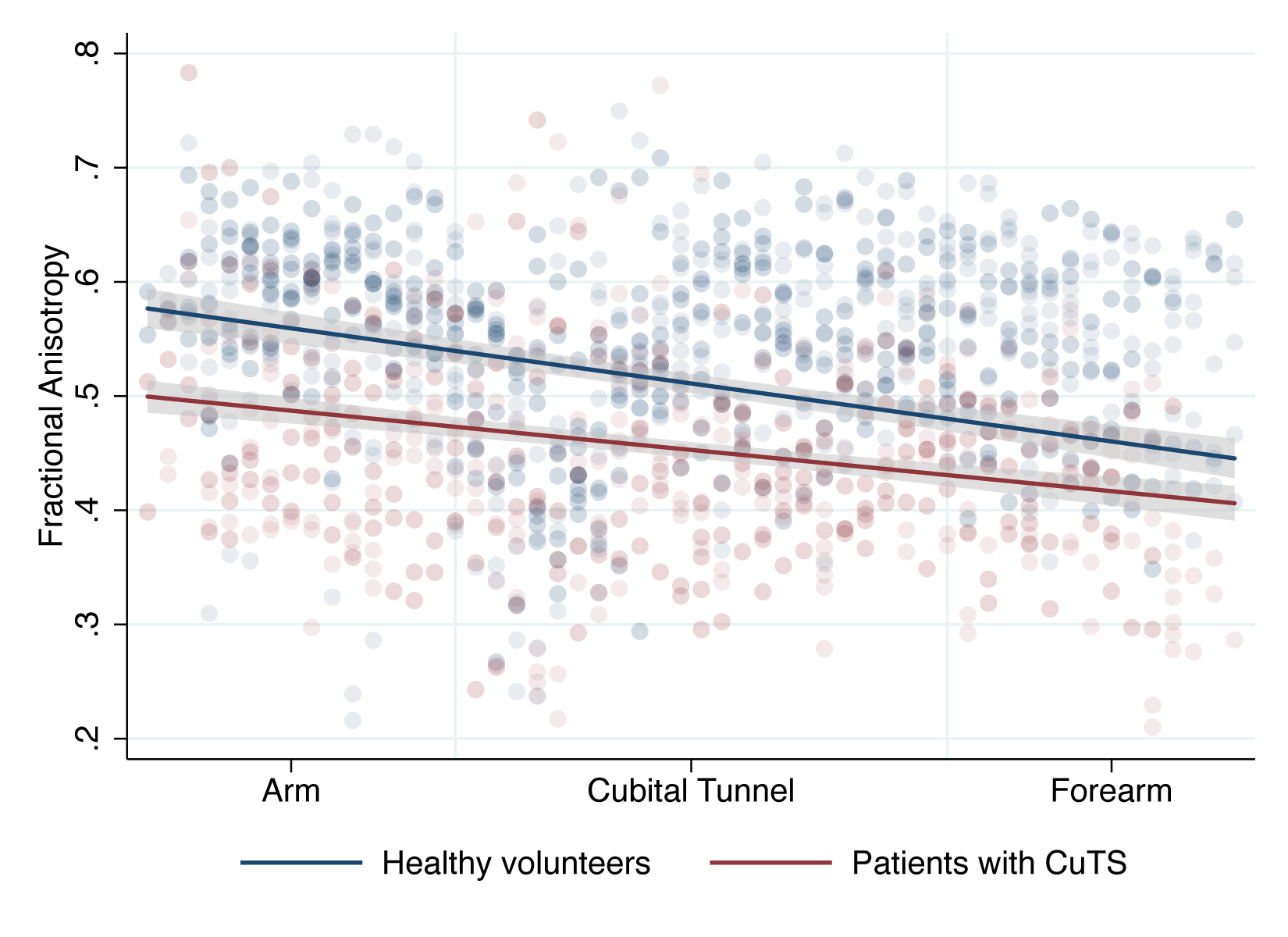
Figure 2. Scatter plot with linear fit (and 95% CI) showing the relationship between Fractional Anisotropy of the ulnar nerve in volunteers and patients, at different positions within the upper limb.