Kirsten Mary Lynch1, Ryan P Cabeen1, Stefanie C Bodison2,3, Arthur W Toga1, and Courtney C.J. Voelker4
1USC Mark and Mary Stevens Institute for Neuroimaging and Informatics, USC Keck School of Medicine, Los Angeles, CA, United States, 2Chan Division of Occupational Science and Occupational Therapy, USC Herman Ostrow School of Dentistry, Los Angeles, CA, United States, 3Division of Pediatrics, USC Keck School of Medicine, Los Angeles, CA, United States, 4USC Caruso Department of Otolaryngology – Head and Neck Surgery, USC Keck School of Medicine, Los Angeles, CA, United States
1USC Mark and Mary Stevens Institute for Neuroimaging and Informatics, USC Keck School of Medicine, Los Angeles, CA, United States, 2Chan Division of Occupational Science and Occupational Therapy, USC Herman Ostrow School of Dentistry, Los Angeles, CA, United States, 3Division of Pediatrics, USC Keck School of Medicine, Los Angeles, CA, United States, 4USC Caruso Department of Otolaryngology – Head and Neck Surgery, USC Keck School of Medicine, Los Angeles, CA, United States
The goal of the
present study was to characterize the regional microstructural development of
the auditory pathway from infancy through adolescence. Our results showed
exponential changes to DTI and NODDI metrics with spatially-varying
developmental trajectories.
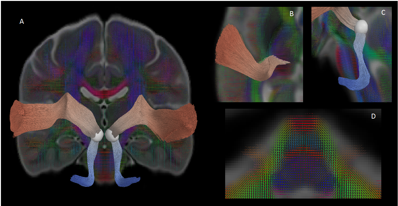
A diffusion MRI atlas used for our auditory pathway reconstruction. The pathway is separated into a lower segment (blue) that connects the cochlea to the inferior colliculus (A, C) and an upper segment that connects the inferior colliculus to the auditory cortex (B). We use multi-compartment diffusion MRI modeling to accurately depict crossing fibers, which are especially important for tractography of brainstem pathways (D).
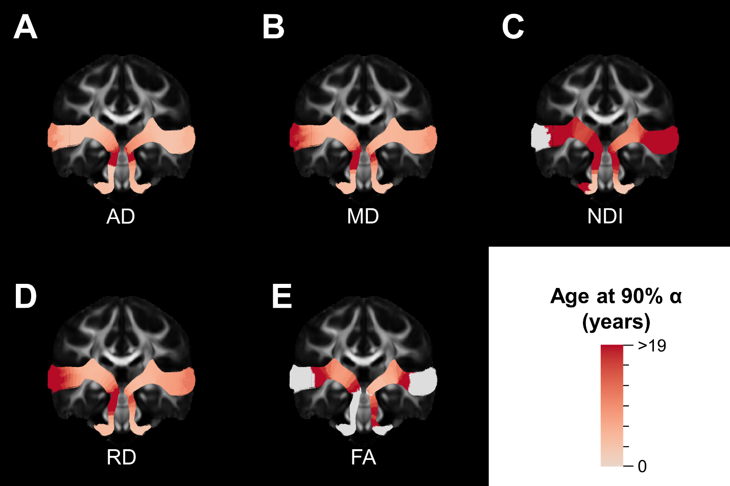
Along-tract
maturation of major white matter tracts. The age at 90% of the asymptotic value
of the fitted growth curve for each point along the auditory pathway are shown
for DTI parameters (AD, RD, MD and FA) and NODDI NDI. Regions shaded in gray
denote points where a growth curve did not provide a good fit.