Yohan van de Looij1,2, Alice Jacquens3,4, Pierre Gressens4,5, Vincent Degos3,4, and Stéphane V Sizonenko1
1Division of Development and Growth, Department of Paediatrics and Gynaecology-Obstetrics, University of Geneva, Geneva, Switzerland, 2Center for Biomedical Imaging, Animal Imaging Technology section, Federal Institute of Technology of Lausanne, Lausanne, Switzerland, 3Department of Anesthesia and Intensive Care, Pitié-Salpêtrière Hospital, Paris, France, 4PROTECT, INSERM, Université Paris Diderot, Sorbonne Paris Cité, Paris, France, 5Centre for the Developing Brain, School of Biomedical Engineering and Imaging Sciences, King's College London, London, United Kingdom
1Division of Development and Growth, Department of Paediatrics and Gynaecology-Obstetrics, University of Geneva, Geneva, Switzerland, 2Center for Biomedical Imaging, Animal Imaging Technology section, Federal Institute of Technology of Lausanne, Lausanne, Switzerland, 3Department of Anesthesia and Intensive Care, Pitié-Salpêtrière Hospital, Paris, France, 4PROTECT, INSERM, Université Paris Diderot, Sorbonne Paris Cité, Paris, France, 5Centre for the Developing Brain, School of Biomedical Engineering and Imaging Sciences, King's College London, London, United Kingdom
Impact-acceleration traumatic brain injury
in the mouse pup brain mimics pediatric brain trauma. By using advanced
diffusion imaging techniques, we were able to accurately characterize white
matter and cortical abnormalities following TBI in the mouse brain.
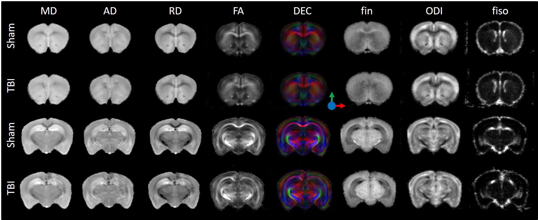
Figure
1: Diffusivity (Mean, MD; Axial, AD and Radial, RD),
fractional anisotropy (FA) and direction encoded color maps (DEC),
intra-neurite volume fraction (fin),
isotropic volume fraction (fiso)
and orientation dispersion index (ODI) at two different image plans for
each group Sham and TBI. Figure represents the average maps over each group.
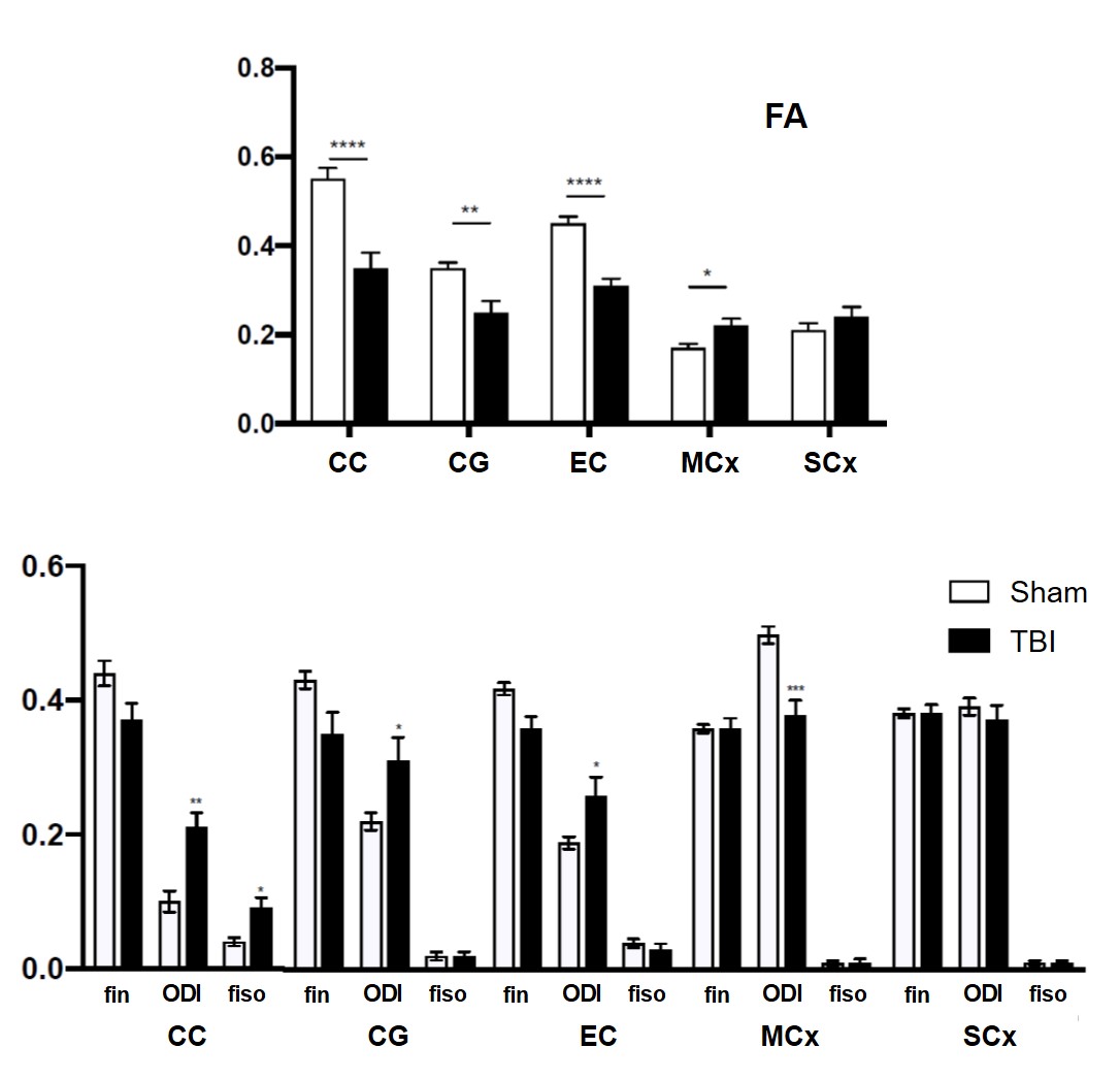
Figure
2: Histogram of mean values ± standard deviation of fractional anisotropy (FA)
(upper case) intra-neurite
volume fraction (fin),
orientation dispersion index (ODI) and isotropic volume fraction (fiso) (lower case) for
Sham and TBI. *P<0.05, **P<0.01, ***P<0.001.