Emily Wheater1, Susan D Shenkin2,3, Susana Muñoz Maniega2,4, Maria Valdés Hernández2,4, Joanna M Wardlaw2,4, Ian J Deary4,5, Mark E Bastin2,4,6, James P Boardman1,2, and Simon R Cox4,5,6
1Centre for Reproductive Health, University of Edinburgh, Edinburgh, United Kingdom, 2Centre for Clinical Brain Sciences, University of Edinburgh, Edinburgh, United Kingdom, 3Geriatric Medicine, Usher Institute, University of Edinburgh, Edinburgh, United Kingdom, 4Lothian Birth Cohorts, University of Edinburgh, Edinburgh, United Kingdom, 5Psychology, University of Edinburgh, Edinburgh, United Kingdom, 6Scottish Imaging Network, A Platform for Scientific Excellence Collaboration (SINAPSE), Edinburgh, United Kingdom
1Centre for Reproductive Health, University of Edinburgh, Edinburgh, United Kingdom, 2Centre for Clinical Brain Sciences, University of Edinburgh, Edinburgh, United Kingdom, 3Geriatric Medicine, Usher Institute, University of Edinburgh, Edinburgh, United Kingdom, 4Lothian Birth Cohorts, University of Edinburgh, Edinburgh, United Kingdom, 5Psychology, University of Edinburgh, Edinburgh, United Kingdom, 6Scottish Imaging Network, A Platform for Scientific Excellence Collaboration (SINAPSE), Edinburgh, United Kingdom
Birth weight is positively associated with
brain tissue volumes at age 73 but not age-associated brain features. Effect of birth weight on brain volumes is
independent of overall body size and is likely to
confer brain tissue reserve in later life.
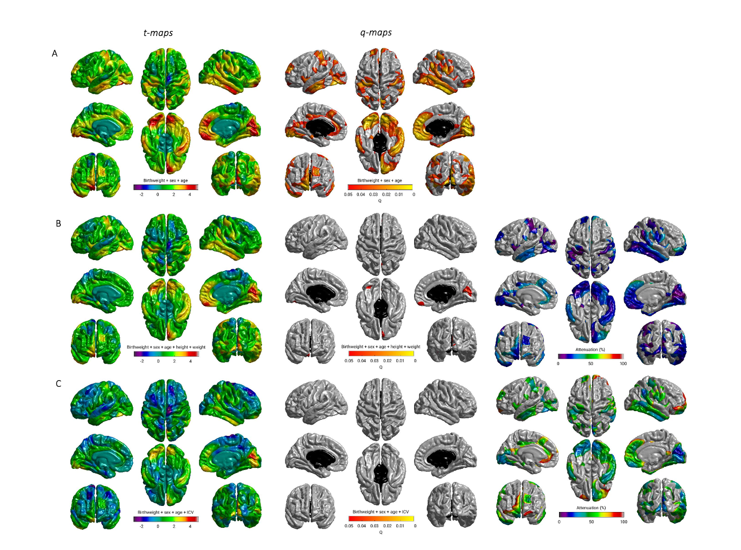
Figure 1. Regional distribution of associations between birth
weight and cortical surface area: A) adjusted for sex and age; B) adjusted for
age, sex, height and weight; C) adjusted for age, sex, and ICV. T maps (left);
FDR q values (middle), far right (B and C) shows the percentage attenuation
between the model shown in A, and the additionally adjusted models shown in B
and C.
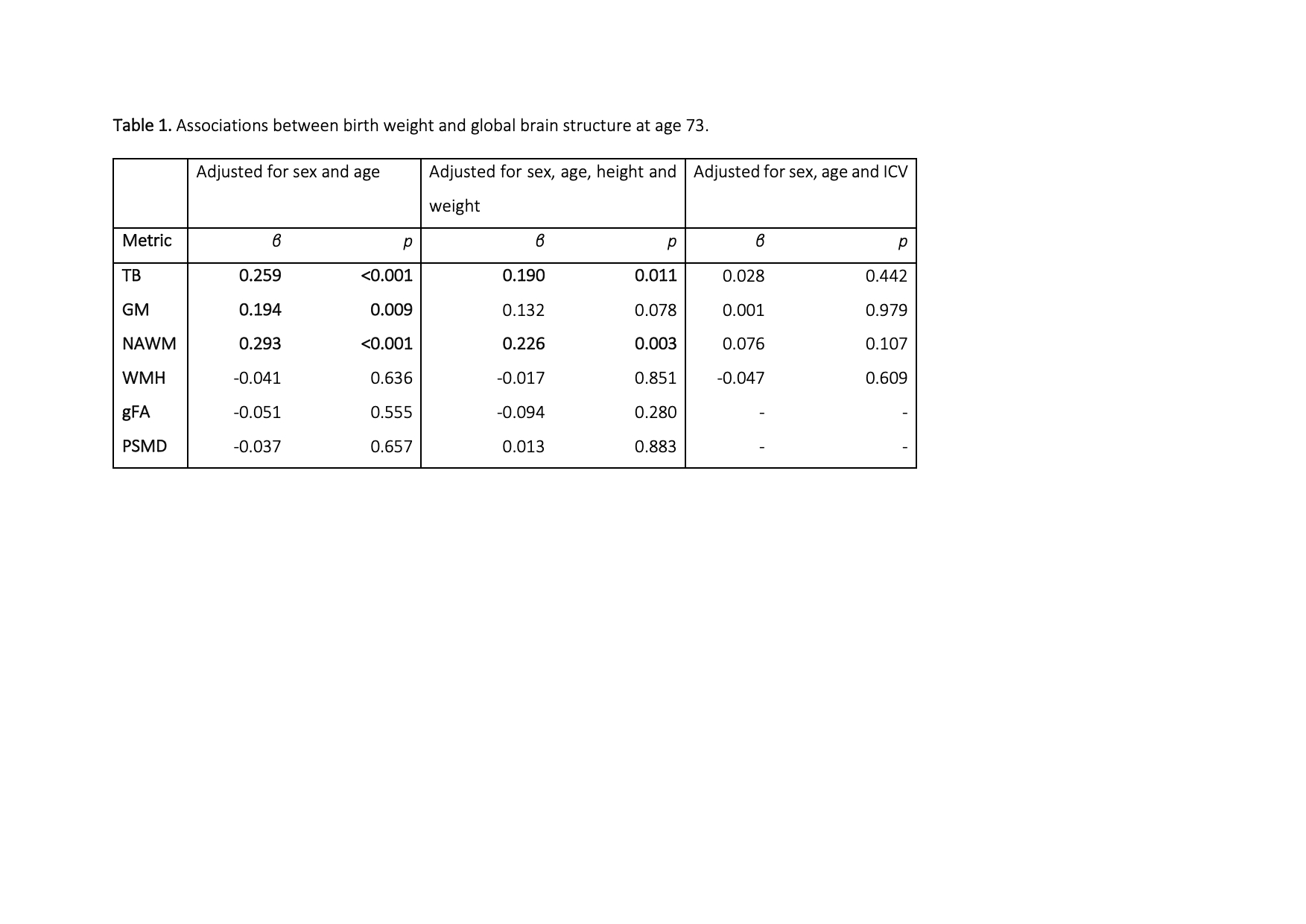
Table 1. Associations between birth weight and global brain structure at
age 73. Standardised regression coefficients between birth weight and
volumetric/white matter microstructure MRI measures in models adjusted for: sex
and age at MRI; sex, age at MRI, height and weight; sex, age at MRI and ICV;
and sex, age at MRI, ICV (intracranial volume), height and weight. Bold
typeface denotes FDR q < 0.05.