Yu-Chieh Jill Kao1, Chia-Feng Lu1, Bao-Yu Hsieh2, Cheng-Yu Chen3, and Chao-Ching Huang4
1National Yang Ming University, Taipei, Taiwan, 2Chang-Gung University, Taoyuan, Taiwan, 3Taipei Medical University, Taipei, Taiwan, 4National Cheng Kung University, Tainan, Taiwan
1National Yang Ming University, Taipei, Taiwan, 2Chang-Gung University, Taoyuan, Taiwan, 3Taipei Medical University, Taipei, Taiwan, 4National Cheng Kung University, Tainan, Taiwan
Temporal and regional profile of ADC-related
MR characteristics early after neonatal hypoxic ischemia between different severity
outcome
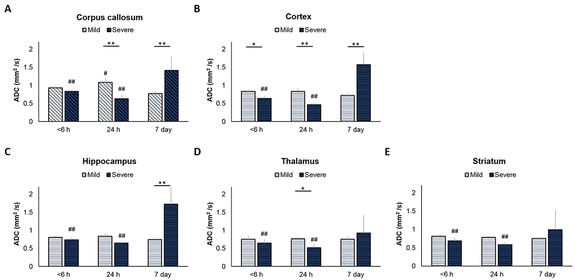
Figure 4
The difference of changes in regional ADC value after HI between the mild and severe
damage outcome groups at each time point after HI. The mean ADC value in the ipsilateral
corpus callosum (A), cortex (B), hippocampus (C), thalamus (D), and striatum (E)
within 6 hours, at 24 hours, and 7 days after HI. The error bars were standard
deviation. *P < 0.05 and **P < 0.005, significant between groups at each
time point. #P < 0.05 and ##P < 0.005, significant from
7 days in the same group.
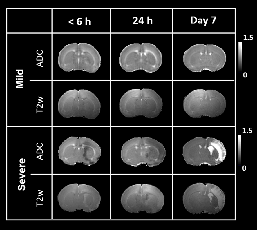
Figure 2
Longitudinal changes in diffusion and T2-weighted MR images at 6 hours, 24
hours, and 7 days after HI. Representative ADC map and T2-weighted images
within 6 hours, at 24 hours, and 7 days after HI in the mild and severe outcome
groups.