Leila Nabulsi1, Katherine E Lawrence1, Vigneshwaran Santhalingam1, Zvart Abaryan1, Christina P Boyle1, Julio E Villalon-Reina1, Talia M Nir1, Iyad Ba Gari1, Alyssa H Zhu1, Elizabeth Haddad1, Alexandra M Muir1, Neda Jahanshad1, and Paul M Thompson1
1Imaging Genetics Center, Mark and Mary Stevens Neuroimaging & Informatics Institute, University of Southern California, Marina del Rey, CA 90292, USA, Los Angeles, CA, United States
1Imaging Genetics Center, Mark and Mary Stevens Neuroimaging & Informatics Institute, University of Southern California, Marina del Rey, CA 90292, USA, Los Angeles, CA, United States
Using the NODDI model of diffusion MRI, we showed that hormone replacement therapy formulation may differentially impact women’s white matter aging trajectories. Duration of therapy, age at onset, and menopause may modulate these effects.
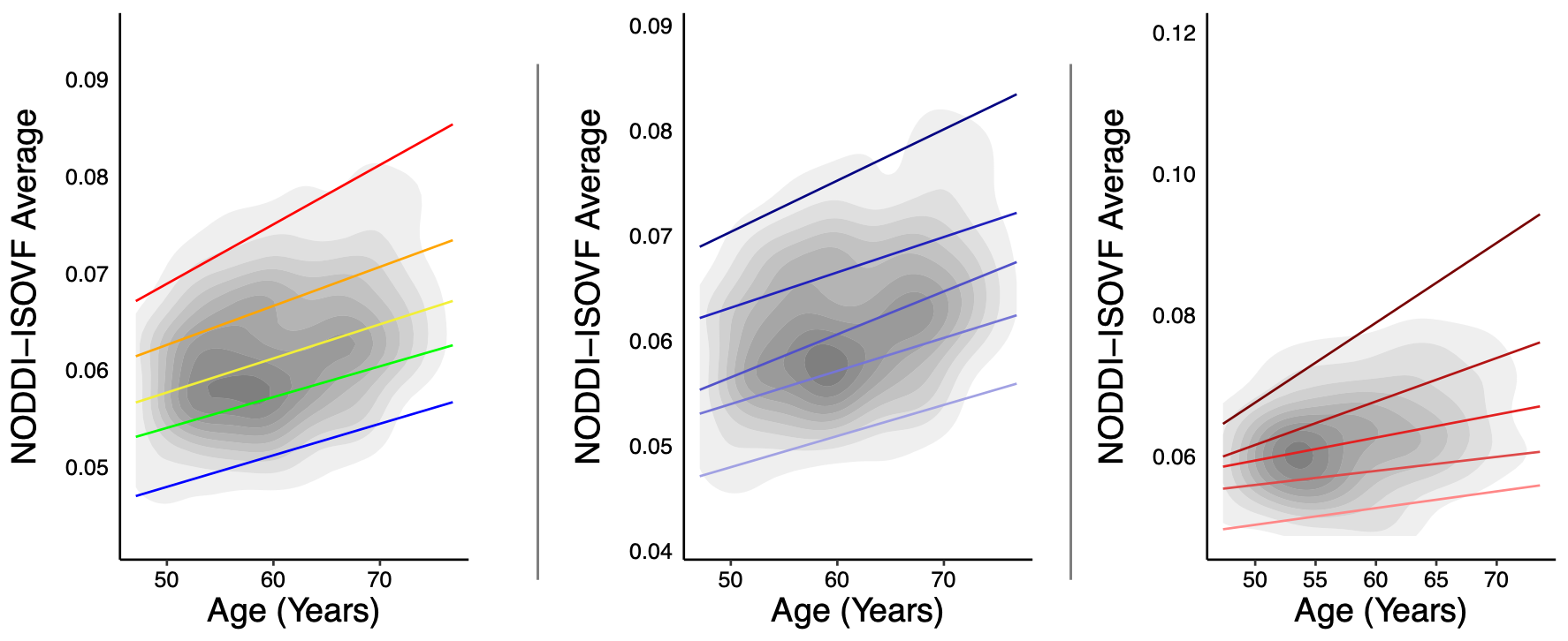
Normative centile reference curves for NODDI-ISOVF, calculated using quantile regression, are presented for estrogen users and combination users, collectively (left panel), and separately for estrogen only (middle panel) and combination (right panel) users. Estrogen-only users displayed a steeper NODDI-ISOVF aging trajectory compared to combination users. Solid colored lines, bottom-up, indicate the following centiles: 5th, 25th, 50th, 75th, 95th. Kernel densities indicating the degree of data point overlap (and sampling density across the age range), are shown in grey.
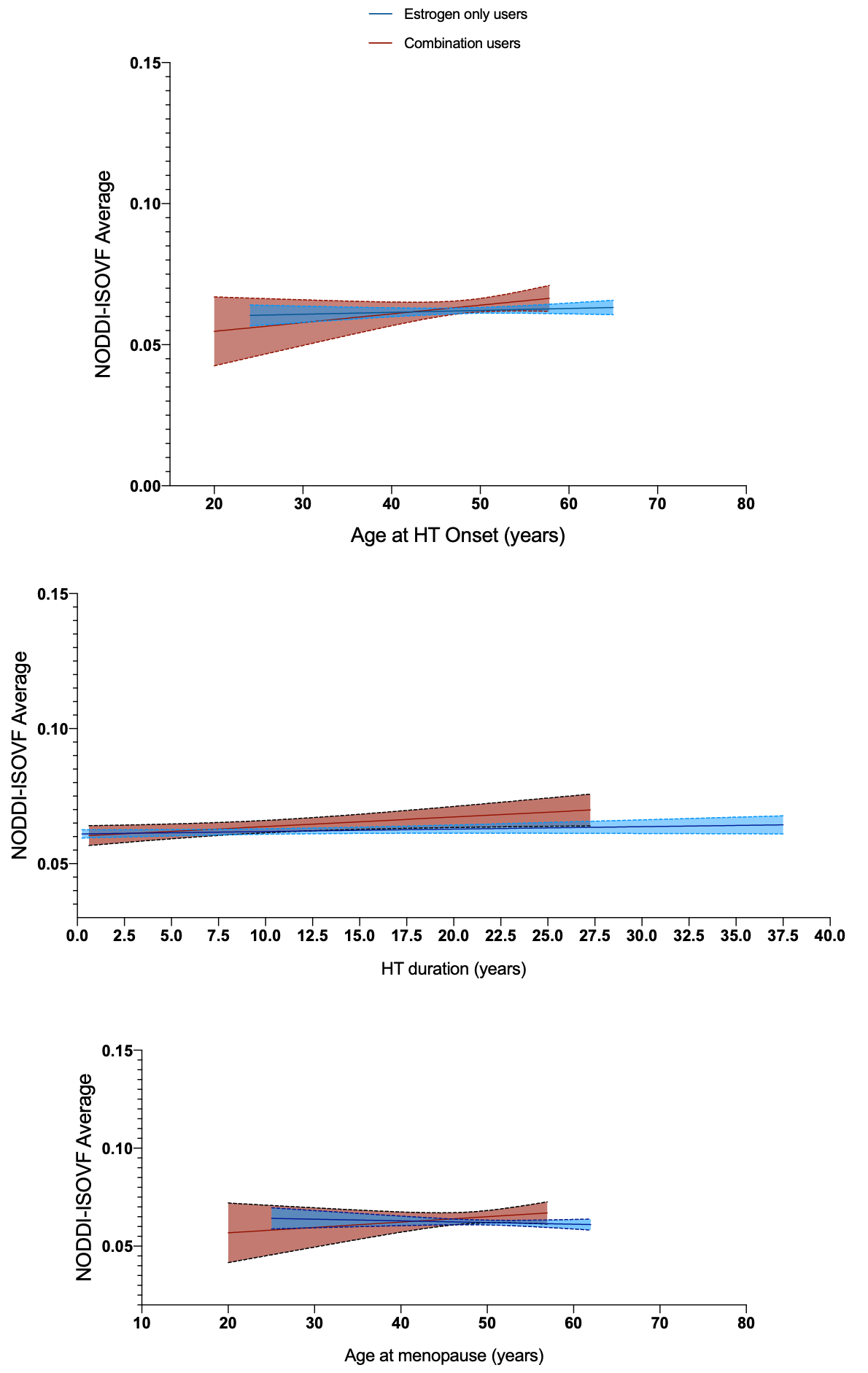
In combination HT (estrogen + progestin) users, later age at HT onset (top panel), prolonged duration of HT therapy (middle panel), and later age at menopause (bottom panel), were associated with higher NODDI-ISOVF, relative to estrogen only HT users. Blue color indicates estrogen only HT users, red indicates combination HT users. Dotted lines represent 95% confidence intervals.