Luke Joel Edwards1, Kerrin J. Pine1, Shubhajit Paul1, Fakhereh Movahedian Attar1, Michael Herbst2, Mirsad Mahmutović3, Boris Keil3, Harald Möller4, Evgeniya Kirilina1,5, and Nikolaus Weiskopf1,6
1Department of Neurophysics, Max Planck Institute for Human Cognitive and Brain Sciences, Leipzig, Germany, 2Gengenbach, Germany, 3Institute of Medical Physics and Radiation Protection, TH Mittelhessen University of Applied Sciences, Giessen, Germany, 4NMR Group, Max Planck Institute for Human Cognitive and Brain Sciences, Leipzig, Germany, 5Center for Cognitive Neuroscience Berlin, Freie Universität Berlin, Berlin, Germany, 6Felix Bloch Institute for Solid State Physics, Leipzig University, Leipzig, Germany
1Department of Neurophysics, Max Planck Institute for Human Cognitive and Brain Sciences, Leipzig, Germany, 2Gengenbach, Germany, 3Institute of Medical Physics and Radiation Protection, TH Mittelhessen University of Applied Sciences, Giessen, Germany, 4NMR Group, Max Planck Institute for Human Cognitive and Brain Sciences, Leipzig, Germany, 5Center for Cognitive Neuroscience Berlin, Freie Universität Berlin, Berlin, Germany, 6Felix Bloch Institute for Solid State Physics, Leipzig University, Leipzig, Germany
A spiral diffusion imaging protocol at 800 µm resolution using a scanner with 300 mT/m gradients and gradient field monitoring shows potential for enhancing the imaging of fine iron-rich regions of the brain.
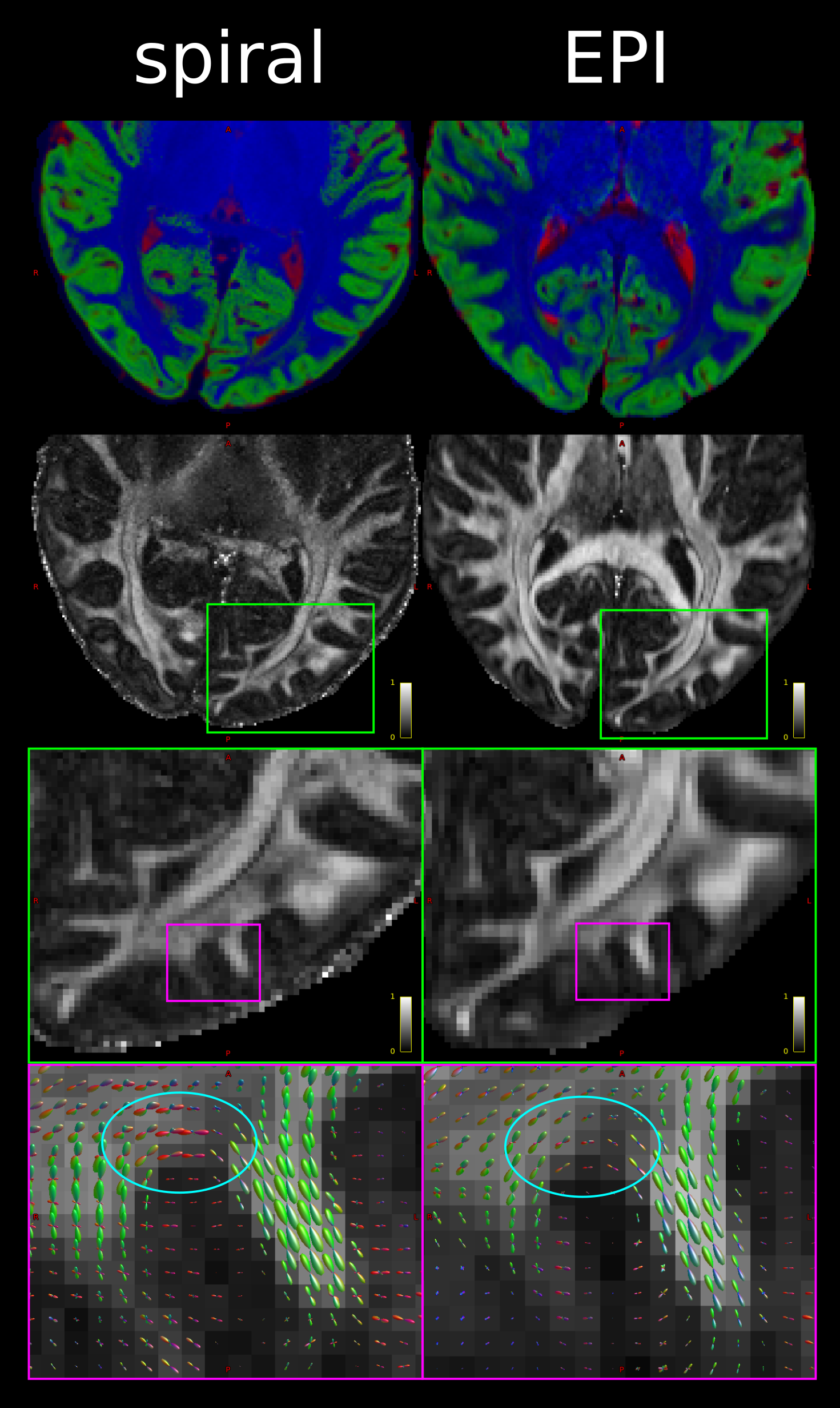
Figure 1: Comparison of spiral and EPI in the occipital lobe. Top: RGB segmentations of DWI data using multi-tissue CSD show better definition of the cortex in the spiral acquisition. Middle: Fractional anistropy (FA) maps further demonstrate the higher effective resolution of the spiral, i.e. less blurring. Bottom: fODFs seem to be more prominent for the iron-rich U-fibres connecting the two gyri (cyan ovals) in the spiral acquisition (short TE) than EPI (long TE). The same glyph scaling was used for both.
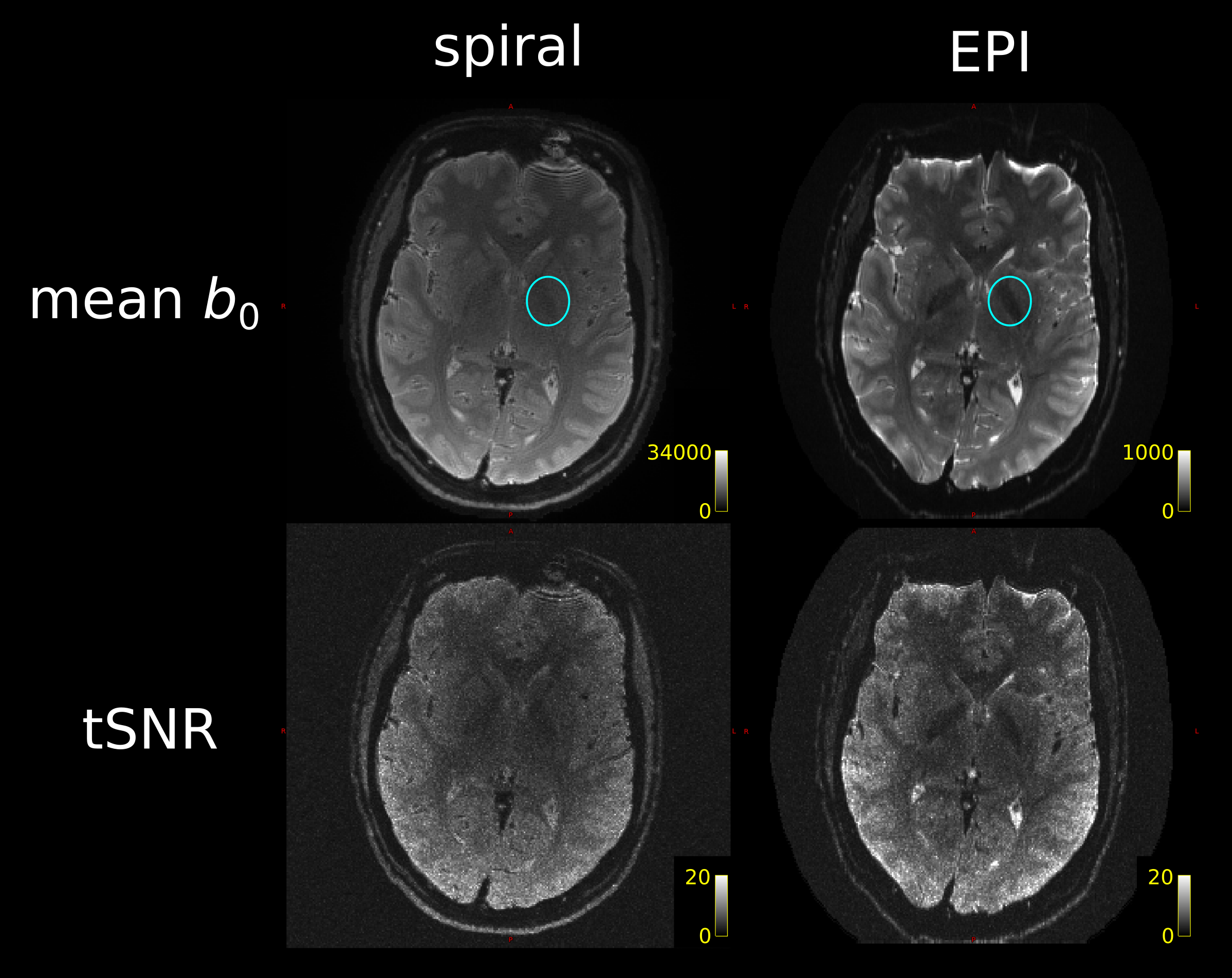
Figure 2: Spiral and EPI data show similar tSNR distributions. The anterior-left (top right of each figure panel) artefact is due to a fiducial oil capsule attached to the subject's forehead. The longer TR of the EPI acquisition resulted in higher cerebrospinal fluid (CSF) signal in the ventricles and near the surface of the brain. The contrast in the putamen (cyan ovals), an iron-rich deep grey matter structure, shows distinct differences between the spiral and EPI because of the different TEs.