Cornelius Eichner1, Michael Paquette1, Guillermo Gallardo1, Christian Bock2, Jenny E. Jaffe3,4, Carsten Jäger1, Evgeniya Kirilina1,5, Ilona Lipp1, Toralf Mildner1, Torsten Schlumm1, Felizitas C Wermter2, Harald E. Möller1, Nikolaus Weiskopf1, Catherine Crockford4,6, Roman Wittig4,6, Angela D Friederici1, and Alfred Anwander1
1Max Planck Institute for Human Cognitive and Brain Sciences, Leipzig, Germany, 2Alfred Wegener Institute Helmholtz Centre for Polar and Marine Research, Bremerhaven, Germany, 3Project Group Epidemiology of Highly Pathogenic Microorganisms, Robert Koch Institute, Berlin, Germany, 4Tai Chimpanzee Project, Centre Suisse de Recherches Scientifiques en Cote d'IVoire, Abidjan, Cote D'ivoire, 5Center for Cognitive Neuroscience Berlin, Freie Universität Berlin, Berlin, Germany, 6Max Planck Institute for Evolutionary Anthropology, Leipzig, Germany
1Max Planck Institute for Human Cognitive and Brain Sciences, Leipzig, Germany, 2Alfred Wegener Institute Helmholtz Centre for Polar and Marine Research, Bremerhaven, Germany, 3Project Group Epidemiology of Highly Pathogenic Microorganisms, Robert Koch Institute, Berlin, Germany, 4Tai Chimpanzee Project, Centre Suisse de Recherches Scientifiques en Cote d'IVoire, Abidjan, Cote D'ivoire, 5Center for Cognitive Neuroscience Berlin, Freie Universität Berlin, Berlin, Germany, 6Max Planck Institute for Evolutionary Anthropology, Leipzig, Germany
Using chimpanzee post-mortem brains
and a comparison between different MRI systems, we showcase the highest
resolution dMRI data yet recorded in chimpanzees. Our research opens the option
of developmental and evolutionary research on structural brain connectivity.
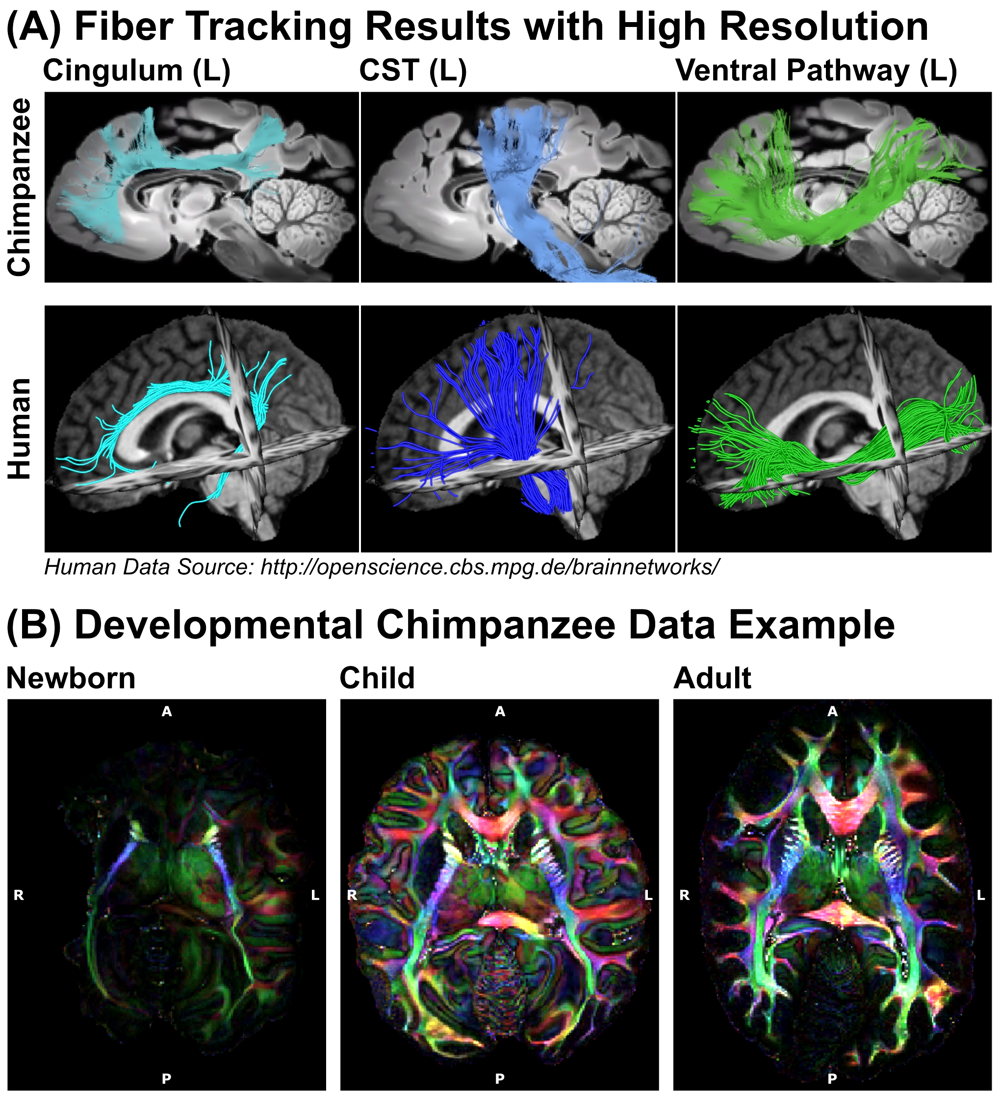
Figure 5: Reconstructions from
Preclinical MRI System - (A) The high-resolution dMRI data, acquired using the preclinical 9.4T MRI, enabled tractography on fine spatial levels. Three respective tract
reconstructions are depicted: Cingulum (turquoise), Corticospinal Tract (blue) and
Ventral Pathway (green) (B) Diffusion data were acquired from various
age groups, enabling a developmental comparison between chimpanzees.
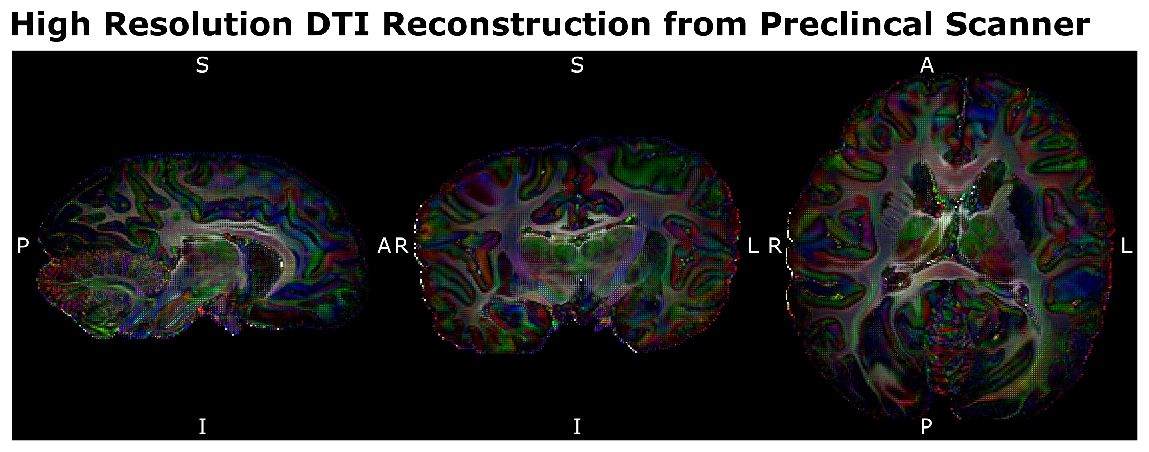
Figure 4: Preclinical Scanner
Acquisition Data Quality - The 500µm isotropic high-resolution dMRI data,
acquired at the preclinical 9.4T MRI system allowed mapping the structural
connectivity of the chimpanzee brain with unprecedented image resolution. (LR –
Left Right, AP – Anterior Posterior, SI – Superior Inferior)