Kulam Najmudeen Magdoom1, Alexandru V. Avram1, Dario Gasbarra2, Qiuyun Fan3, Thomas Witzel3, Susie Y Huang3, and Peter J Basser1
1National Institute of Health, Bethesda, MD, United States, 2University of Helsinki, Helsinki, Finland, 3Massachusetts General Hospital and Harvard Medical School, Charlestown, MA, United States
1National Institute of Health, Bethesda, MD, United States, 2University of Helsinki, Helsinki, Finland, 3Massachusetts General Hospital and Harvard Medical School, Charlestown, MA, United States
A new experimental design and analysis technique is introduced to make an unbiased estimate of mean and covariance tensors of diffusion tensor distribution. Applying it in-vivo on a human brain revealed new information about brain microstructure.
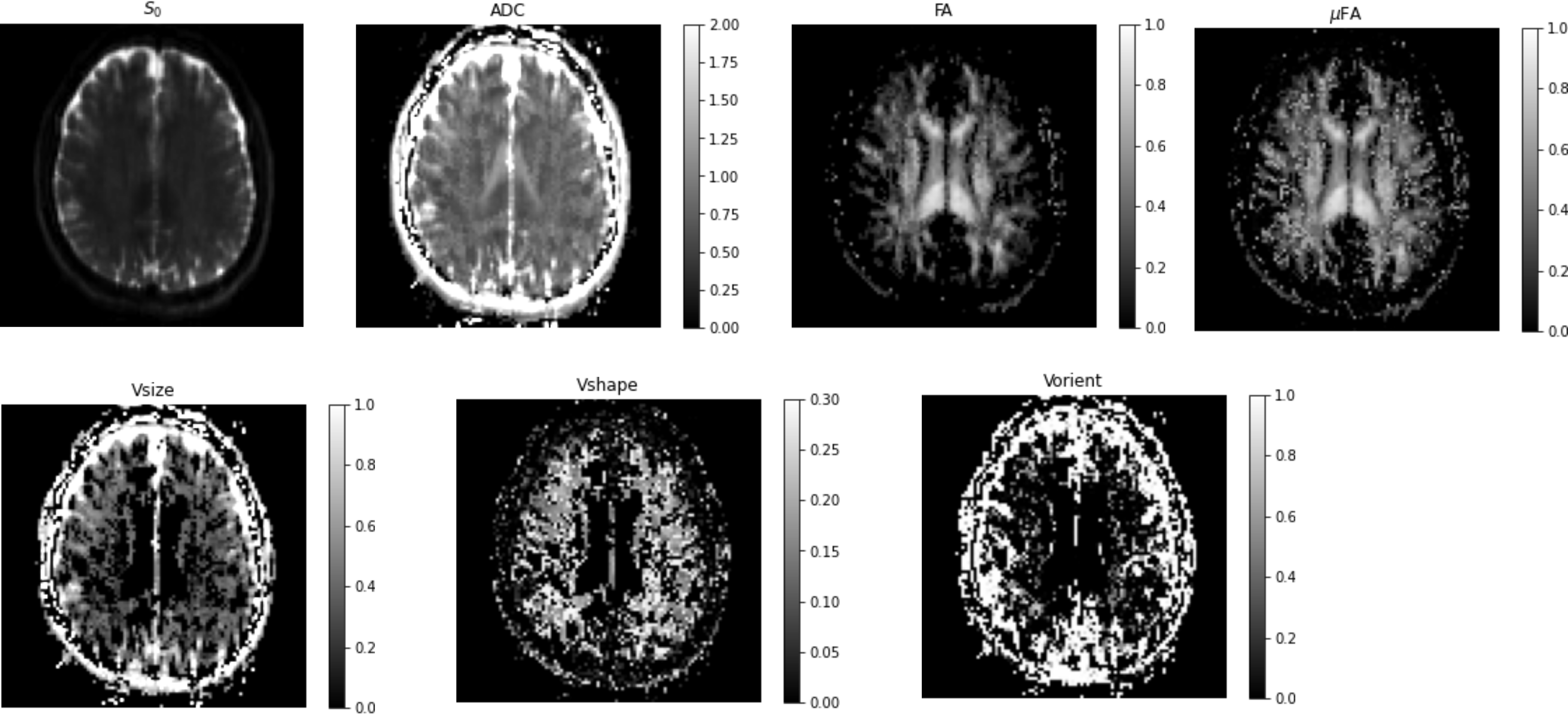
Figure 4: DTD results with the brain showing the estimated parametric maps. S0 – Non-diffusion weighted MRI, FA - fractional anisotropy map, μFA - microscopic FA map, ADC - apparent diffusion coefficient, Vsize, Vshape, Vorient – Size, shape and orientation heterogeneity metrics. The units for ADC and Vsize are in μm2/ms.
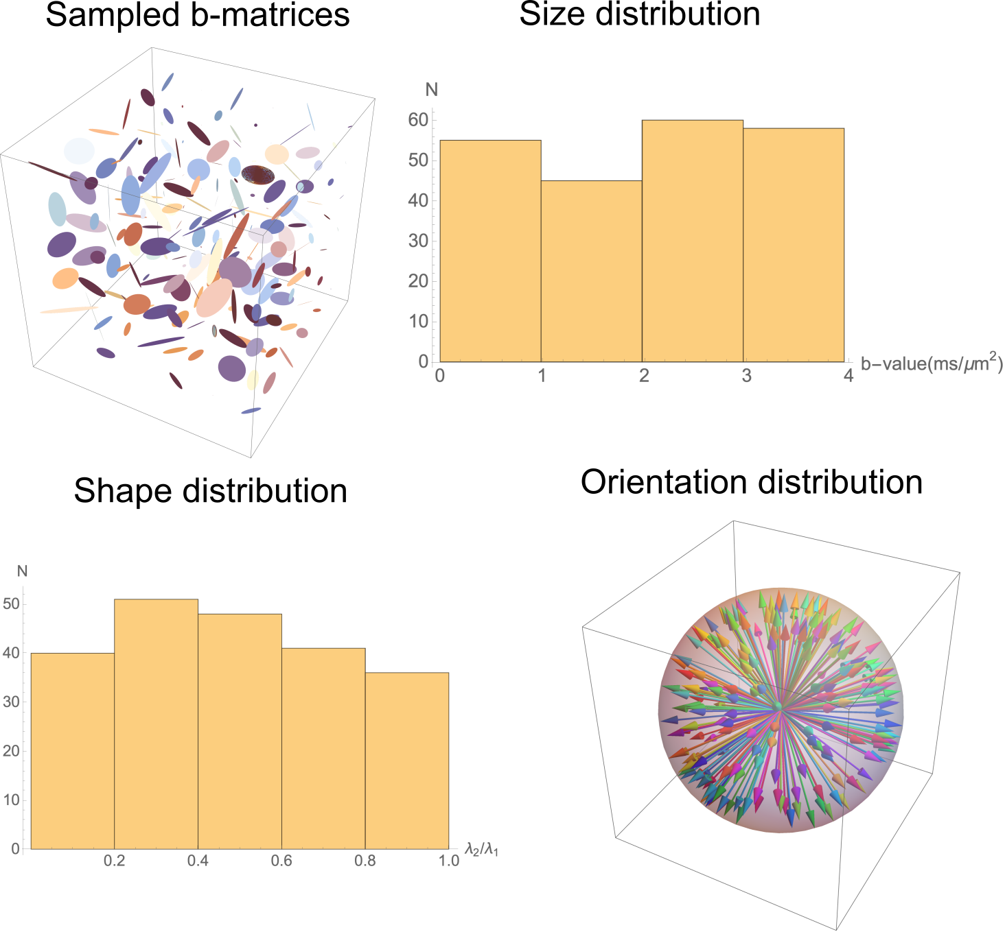
Figure 2: Experimental design showing the prescribed b-matrices shown using ellipsoids (top left), and the distribution of b-values (top right), shapes characterized by ratio of the two non-zero eigenvalues of rank-2 b-matrix (bottom left) and orientation dispersion (bottom right) they produce obtained by random sampling.