Rui Zeng1, Jinglei Lv2, He Wang3, Luping Zhou2, Michael Barnett2, Fernando Calamante2, and Chenyu Wang2
1School of Biomedical Engineering, The University of Sydney, Sydney, Australia, 2The University of Sydney, Sydney, Australia, 3Fudan University, Shanghai, China
1School of Biomedical Engineering, The University of Sydney, Sydney, Australia, 2The University of Sydney, Sydney, Australia, 3Fudan University, Shanghai, China
A deep learning model called FODSRM was developed for fiber
orientation distribution (FOD) super-resolution, which enhances single-shell
FOD computed from clinic-quality dMRI data to obtain the
super-resolved quality that would have been produced
from advanced research scanners.
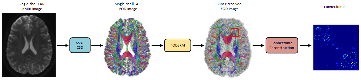
Figure 1. An
overview of the pipeline for applying FODSRM in connectome reconstruction. A
given single-shell low-angular-resolution (LAR) dMRI image is first processed
by single-shell 3-tissue CSD method to generate the single-shell LAR FOD image,
which is then taken as input by FODSRM to generate the super-resovled FOD
image. The super-resolved FOD image is used for reliable connectome reconstruction.
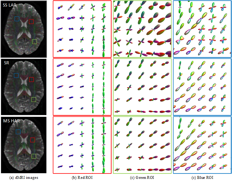
Figure 2. Illustration of FOD super
resolution. Our model is able to generate high-angular-resolution (HAR) FOD
images by enhancing the corresponding low-angular-resolution (LAR) FOD images.
The LAR FOD images are obtained with SS3T-CSD on single-shell
low-angular-resolution (typically around 32 gradient directions) data, which
are extensively used for clinical purposes. The recovered HAR FOD images have
comparable quality to those from MSMT-CSD. Three representative zoomed regions
(red, green and blue) are shown.