Maria Eugenia Caligiuri1, Andrea Quattrone2, Alessandro Mechelli2, and Aldo Quattrone1
1Neuroscience Research Center, University "Magna Graecia", Catanzaro, Italy, 2Institute of Neurology, University "Magna Graecia", Catanzaro, Italy
1Neuroscience Research Center, University "Magna Graecia", Catanzaro, Italy, 2Institute of Neurology, University "Magna Graecia", Catanzaro, Italy
Altered PDD orientation in the splenium underlies iNPH but not PSP or AD. Splenium fibers might be damaged in PSP and AD due to Wallerian degeneration, while in iNPH ventricles could "push" the bundle upwards, deviating the physiological laterolateral PDD of the callosal fibers.
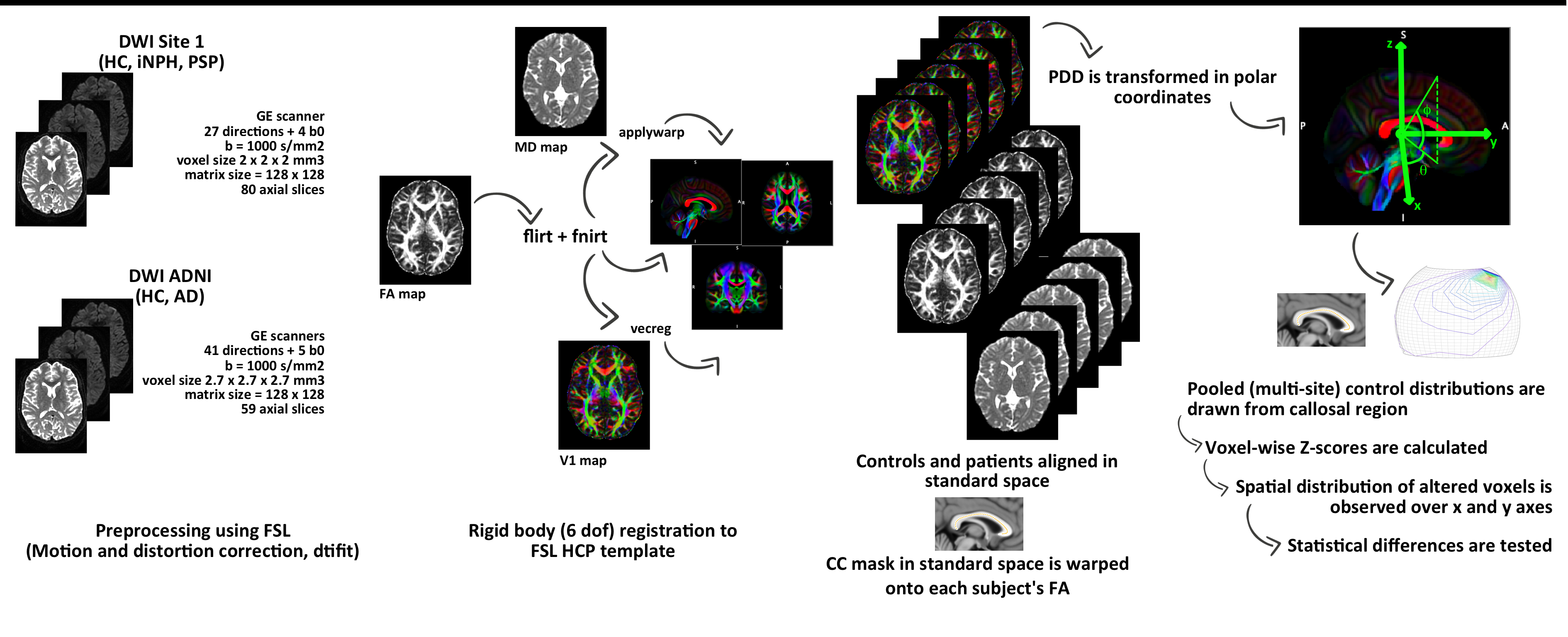
Figure 1: Image processing and analysis workflow.
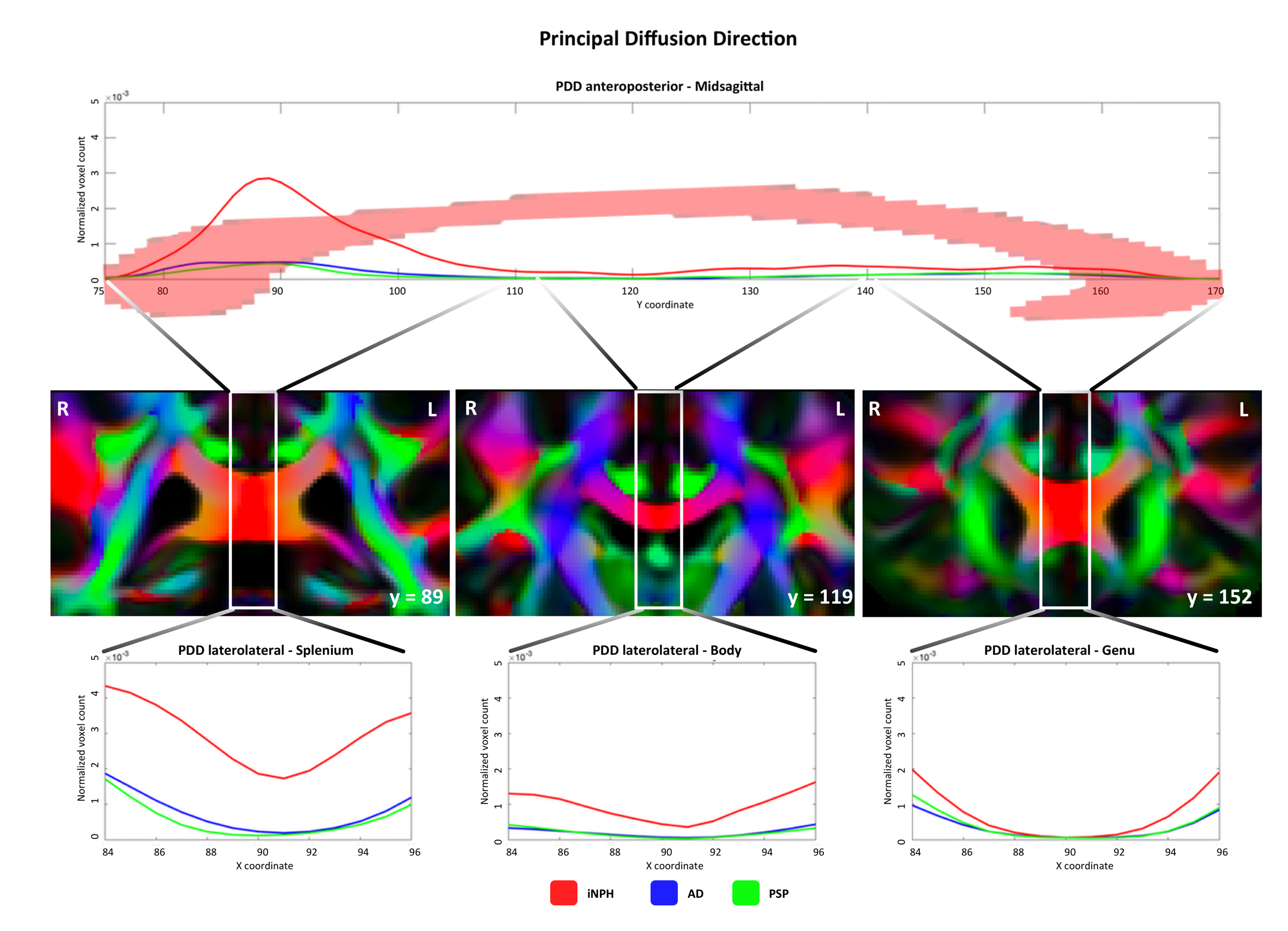
Figure 2. Top row: altered PDD voxel count compared to control distribution along the anteroposterior axis (z-scores for both theta and phi values > 2.5 or < -2.5). Middle row: CC splenium, body and genu in the coronal plane of PDD-color-coded FA template. Bottom row: altered PDD voxel count along the latero-lateral axis in CC subregions. Abbreviations: PDD = Principal Diffusion Direction; FA = Fractional Anisotropy; iNPH = idiopathic Normal Pressure Hydrocephalus; AD = Alzheimer’s Disease; PSP = Progressive Supranuclear Palsy.