Ayhan Gursan1, Martijn Froeling1, Arjan D. Hendriks1, Dimitri Welting1, Dennis W.J. Klomp1, and Jeanine J. Prompers1
1Department of Radiology, University Medical Center Utrecht, Utrecht, Netherlands
1Department of Radiology, University Medical Center Utrecht, Utrecht, Netherlands
Anisotropic
motional averaging in skeletal muscle leads to deuterium quadrupole moment
splittings of the HDO signal, the size of which depends on the angle between
the muscle fibers and B0.
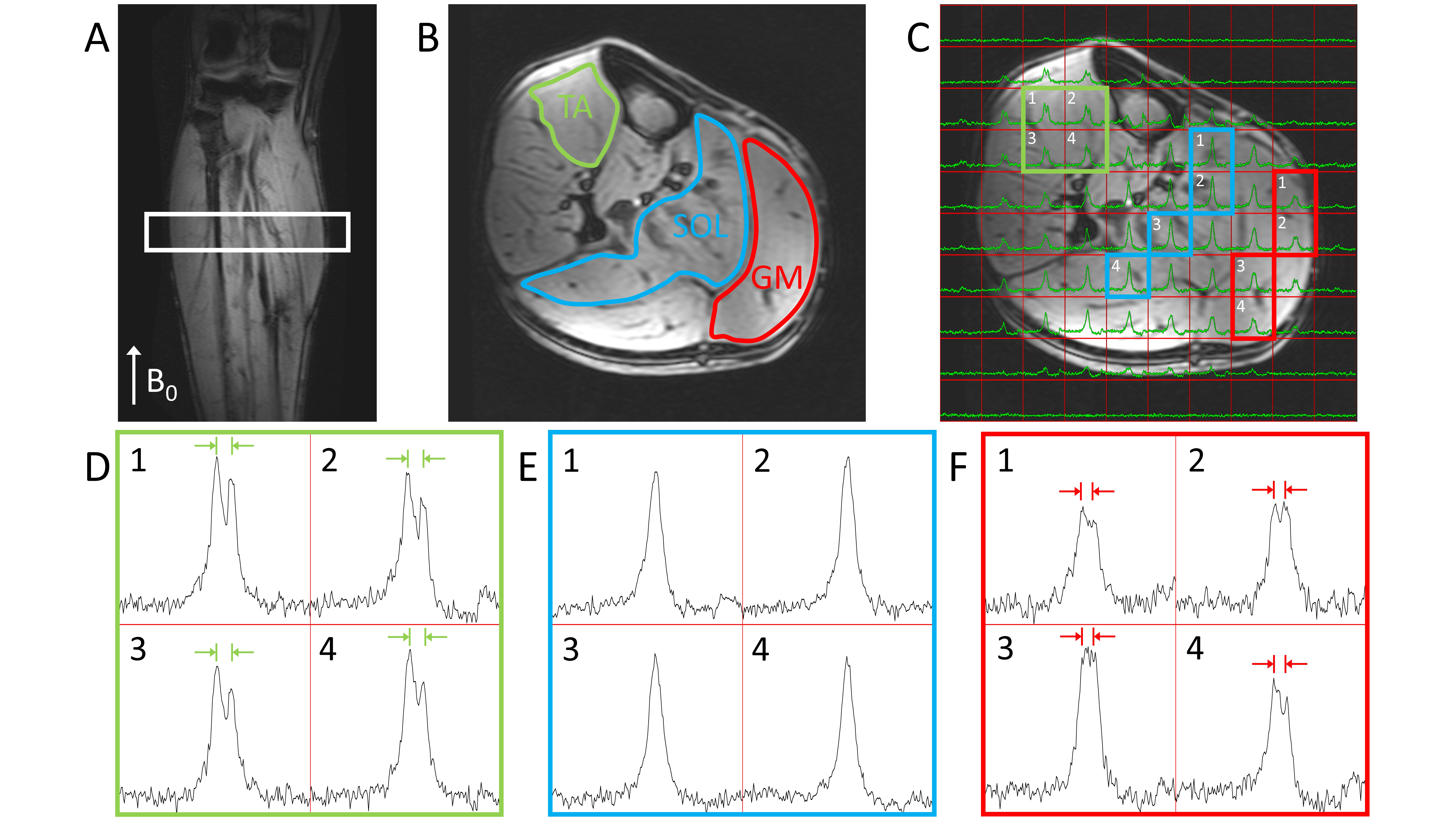
Figure 1. Natural abundance DMRSI data in the right lower leg positioned
parallel with B0. (A) Coronal T1w image. The white rectangle
indicates the slice shown in panels B and C. Transversal T1w image with (B)
segmentation of Tibialis Anterior (TA), Soleus (SOL) and Gastrocnemius Medialis
(GM) muscles, and (C) overlaid with DMRSI data. Spectra from selected voxels in
TA (D), SOL (E) and GM (F), as indicated in panel C, showing the signal from
HDO at 4.7 ppm (x-axis range: 0-10 ppm = 457 Hz).
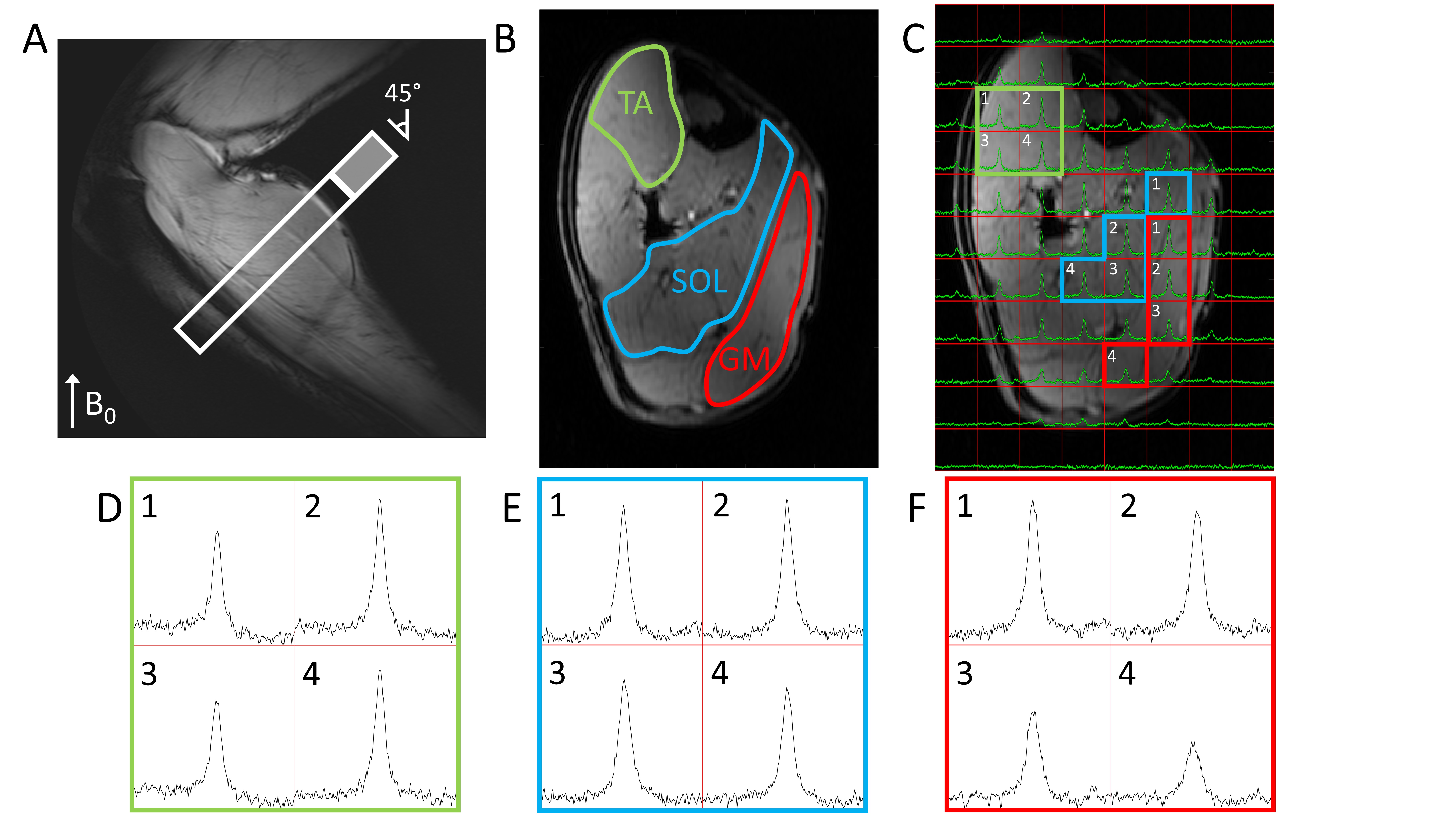
Figure 3. Natural abundance DMRSI data in the right lower leg positioned at an angle of approximately 45° with respect to B0. (A) Sagittal T1w image. The white rectangle indicates the slice shown in panels B and C (cropped in AP direction to size of open part of rectangle). Transversal T1w image with (B) segmentation of TA, SOL and GM muscles, and (C) overlaid with DMRSI data. Spectra from selected voxels in TA (D), SOL (E) and GM (F), as indicated in panel C, showing the signal from HDO at 4.7 ppm (x-axis range: 0-10 ppm = 457 Hz).