Dennis Kleimaier1, Simon Reichert1, Victor Schepkin2, and Lothar R. Schad1
1Computer Assisted Clinical Medicine, Heidelberg University, Mannheim, Germany, 2National High Magnetic Field Laboratory, Florida State University, Tallahassee, FL, United States
1Computer Assisted Clinical Medicine, Heidelberg University, Mannheim, Germany, 2National High Magnetic Field Laboratory, Florida State University, Tallahassee, FL, United States
Glutamic acid, arginine, and lysine as well as the small sized protein α-LA
resulted only in a significant reduction in the sodium relaxation times without
a detectable sodium TQ signal. In contrast, a BSA concentration of 3% w/v was
already sufficient to yield a well detected sodium TQ signal.
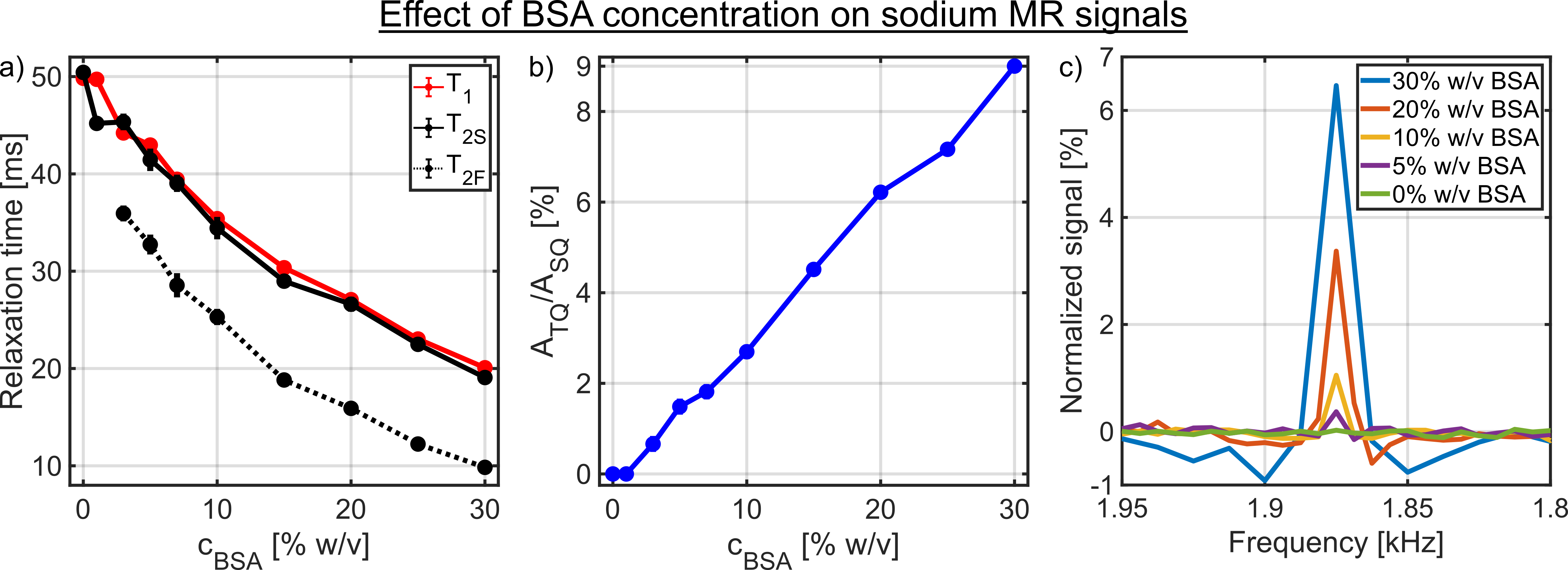
Fig.4: a) Dependence of the longitudinal and the transverse relaxation times on
the BSA concentration. In the samples with a BSA concentration of 0% and 1%
w/v, no sodium TQ signal was observed. Consequently, the TQTPPI FID was only fitted by
a mono-exponential SQ signal decay. B) Dependence of the sodium TQ signal on
the BSA concentration. The sodium TQ signal increased almost linearly with the
BSA concentration. c) Zoomed TQTPPI spectra for different BSA concentrations.
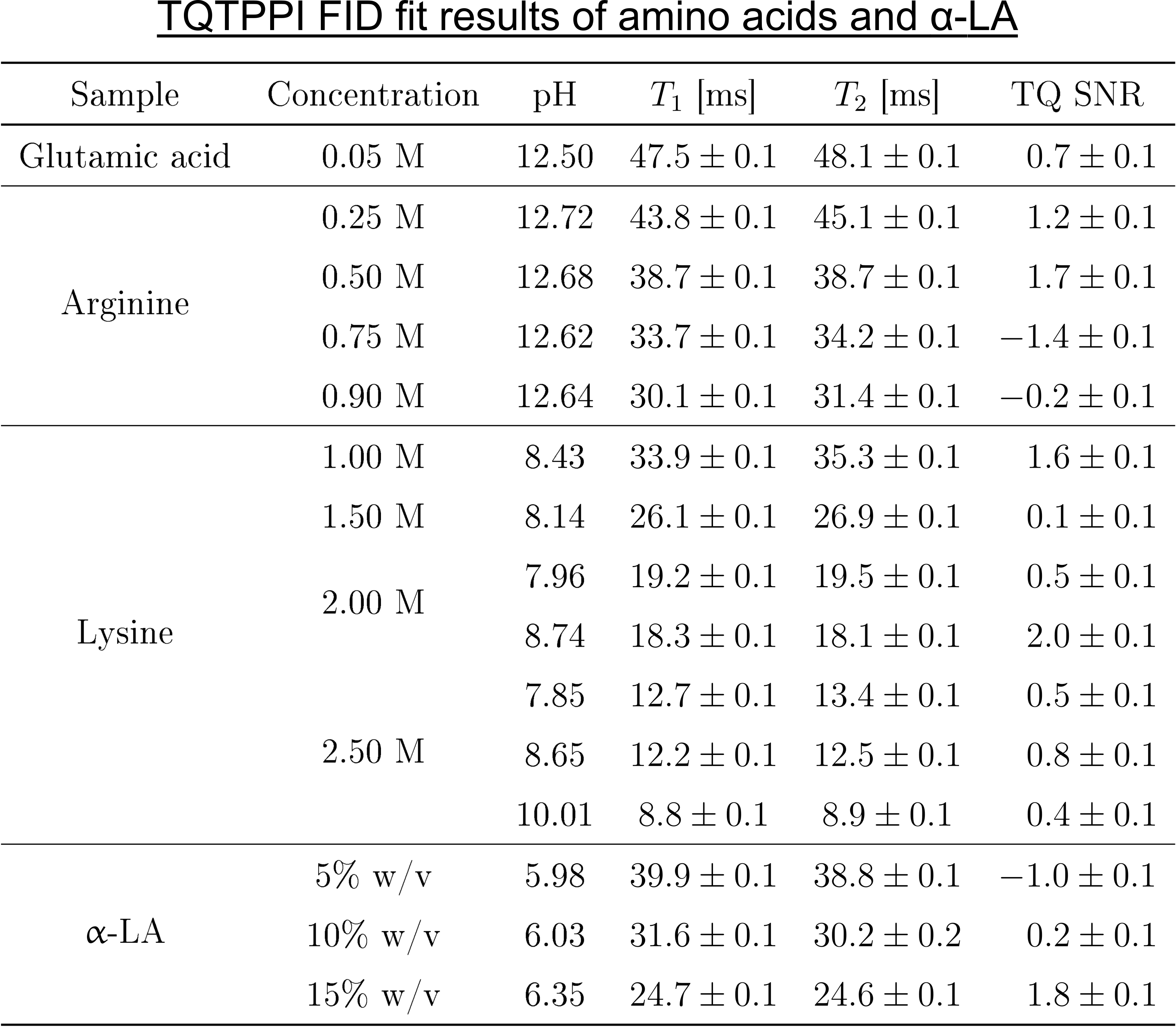
Fig.2: TQTPPI FID fit results of the
samples containing amino acids or α-LA.
The TQ SNR of each sample was less than three and thus the TQ term in the
TQTPPI FID fit function was omitted. Hence, the TQTPPI FID fit function
consisted only of a mono-exponential SQ signal decay. Exemplary TQTPPI spectra
of these samples are shown in Fig.3.