Hao Song1, Burak Akin1, Johannes Fischer1, Ali Caglar Özen1,2, Stefan Schumann3, and Michael Bock1,2
1Dept. of Radiology, Medical Physics, Medical Center University of Freiburg, Faculty of Medicine, University of Freiburg, Freiburg, Germany, 2German Consortium for Translational Cancer Research (DKTK), Partner Site Freiburg, Freiburg, Germany, 3Dept. of Anesthesiology and Critical Care, Medical Center University of Freiburg, Faculty of Medicine, University of Freiburg, Freiburg, Germany
1Dept. of Radiology, Medical Physics, Medical Center University of Freiburg, Faculty of Medicine, University of Freiburg, Freiburg, Germany, 2German Consortium for Translational Cancer Research (DKTK), Partner Site Freiburg, Freiburg, Germany, 3Dept. of Anesthesiology and Critical Care, Medical Center University of Freiburg, Faculty of Medicine, University of Freiburg, Freiburg, Germany
The CMRO2 values were determined in brain
subcortical structures including thalamus, dorsal striatum, caudate nucleus and
insula cortex using dynamic 17O-MRI.
Our
results show the feasibility of measuring local CMRO2 in brain subcortical
structures using 17O-MRI at 3T.
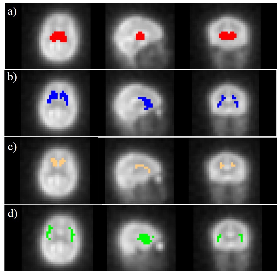
Figure 1. Co-registered masks of ROIs including thalamus (a), dorsal striatum (b), caudate nucleus (c) and
insula (d) superimposed on the averaged 17O MR images.
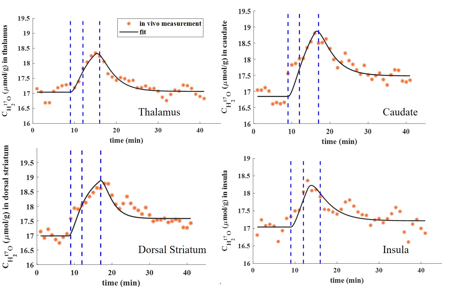
Figure 2. Dynamic H217O concentration (CH217O) in all examined ROIs. Blue dashed lines indicate the 17O2 inhalation time period.