Josephine L Tan1,2, Daniel Djayakarsana1,2, Hanzhi Wang1,2, Rachel W Chan2, Colleen Bailey1,2, and Angus Z Lau1,2
1Medical Biophysics, University of Toronto, Toronto, ON, Canada, 2Physical Sciences, Sunnybrook Research Institute, Toronto, ON, Canada
1Medical Biophysics, University of Toronto, Toronto, ON, Canada, 2Physical Sciences, Sunnybrook Research Institute, Toronto, ON, Canada
Significant decreases in 2H-labeled lactate signal were detected by deuterium magnetic resonance spectroscopy in acute myeloid leukemia cells 48 hours after cisplatin treatment, and significantly correlated with apoptotic cell death and extracellular lactate concentration.
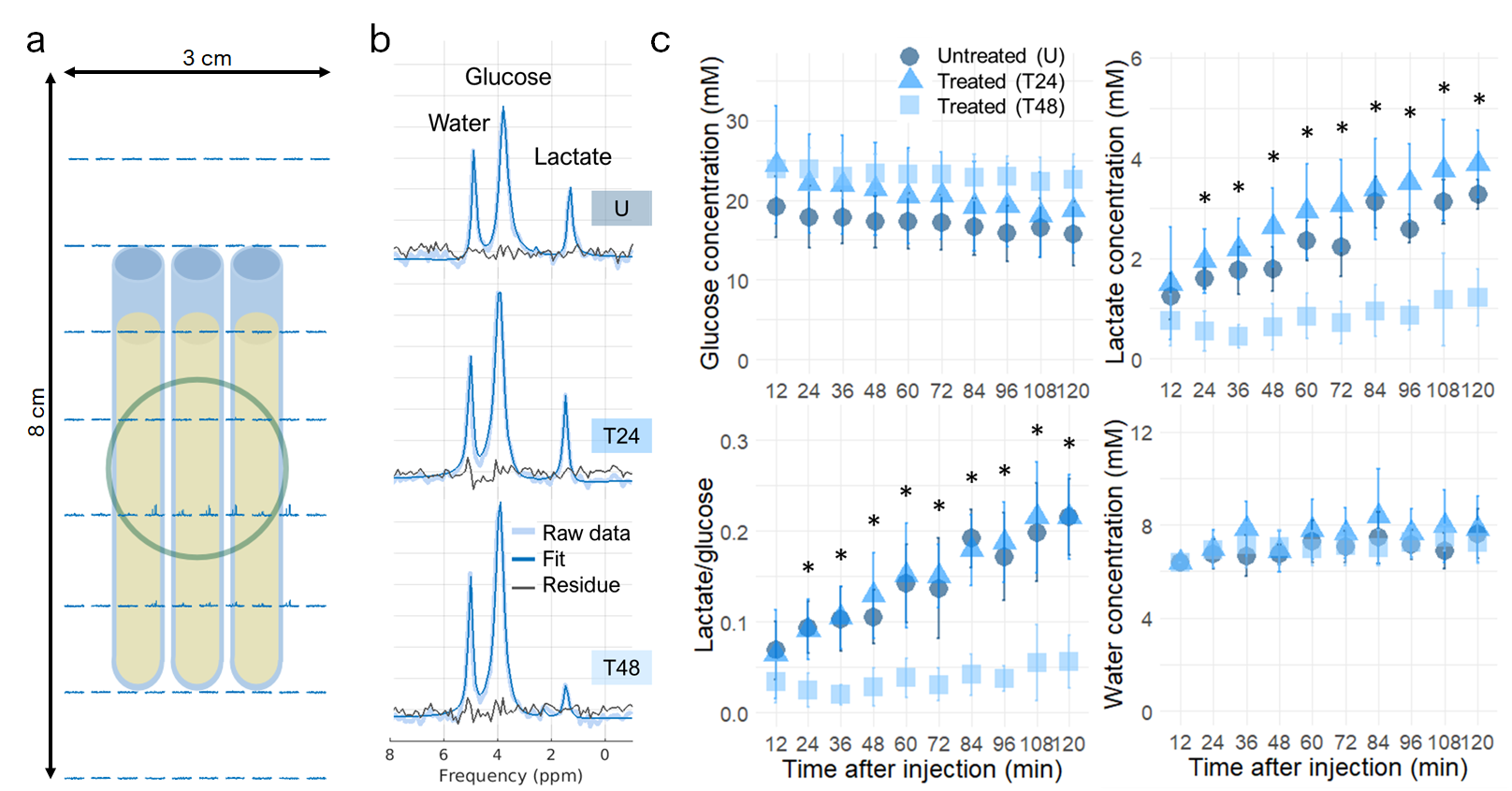
(a) Experimental setup consisting of three AML sample tubes lying on top of the 2H coil (green circle), with overlaid 2H spectra. (b) Single voxel 2H spectra for the untreated (U) and cisplatin-treated samples (T24 and T48) two hours after injection of [6,6’-2H2]glucose. (c) Time course of 2H-labeled species 10 minutes after injection of [6,6’-2H2]glucose. Error bars are the standard deviation across 6 samples. Stars indicate a statistically significance difference between T48 and U, and T48 and T24 (p<0.05)
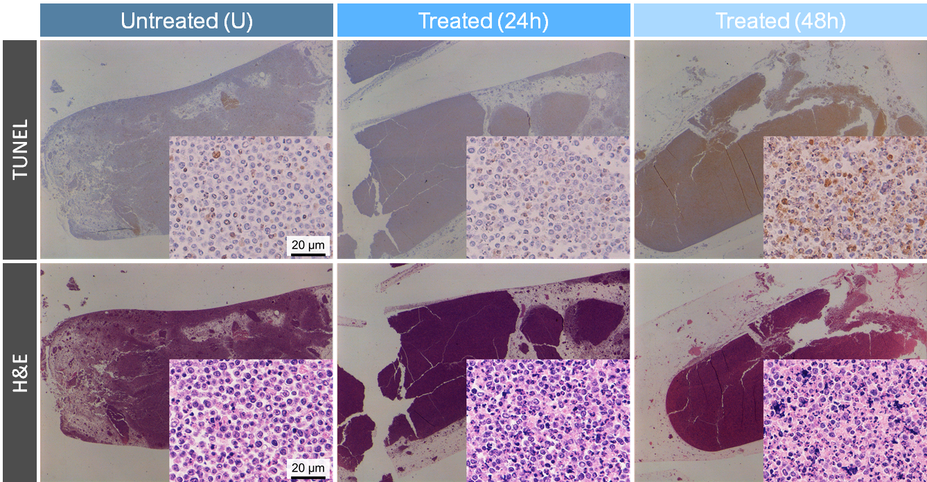
Representative TUNEL and H&E stained sections (10x and 200x magnification) from untreated and cisplatin-treated cell pellets, which were fixed in formalin for at least 96 hours after scanning. Formalin fixation may have caused pellet size shrinkage.