Loreen Ruhm1,2, Nikolai Avdievitch1, Theresia Ziegs1,2, Armin M. Nagel3,4, Henk M. De Feyter5, Robin A. de Graaf5, and Anke Henning1,6
1High-Field MR Center, Max Planck Institute for Biological Cybernetics, Tuebingen, Germany, 2IMPRS for Cognitive and Systems Neuroscience, Eberhard-Karls University, Tuebingen, Germany, 3Institute of Radiology, University Hospital Erlangen, Erlangen, Germany, 4Division of Medical Physics in Radiology, German Cancer Research Center (DKFZ), Heidelberg, Germany, 5Radiology and Biomedical Imaging, Yale University, New Haven, CT, United States, 6Advanced Imaging Research Center, UT Southwestern Medical Center, Dallas, TX, United States
1High-Field MR Center, Max Planck Institute for Biological Cybernetics, Tuebingen, Germany, 2IMPRS for Cognitive and Systems Neuroscience, Eberhard-Karls University, Tuebingen, Germany, 3Institute of Radiology, University Hospital Erlangen, Erlangen, Germany, 4Division of Medical Physics in Radiology, German Cancer Research Center (DKFZ), Heidelberg, Germany, 5Radiology and Biomedical Imaging, Yale University, New Haven, CT, United States, 6Advanced Imaging Research Center, UT Southwestern Medical Center, Dallas, TX, United States
We present first DMI data of the
human brain at B0 = 9.4T with a high spatial resolution acquired at
a Siemens whole body scanner with a dedicated coil design. We investigated the
dynamic uptake of [6,6’-2H]-glucose after oral administration in
different brain areas.
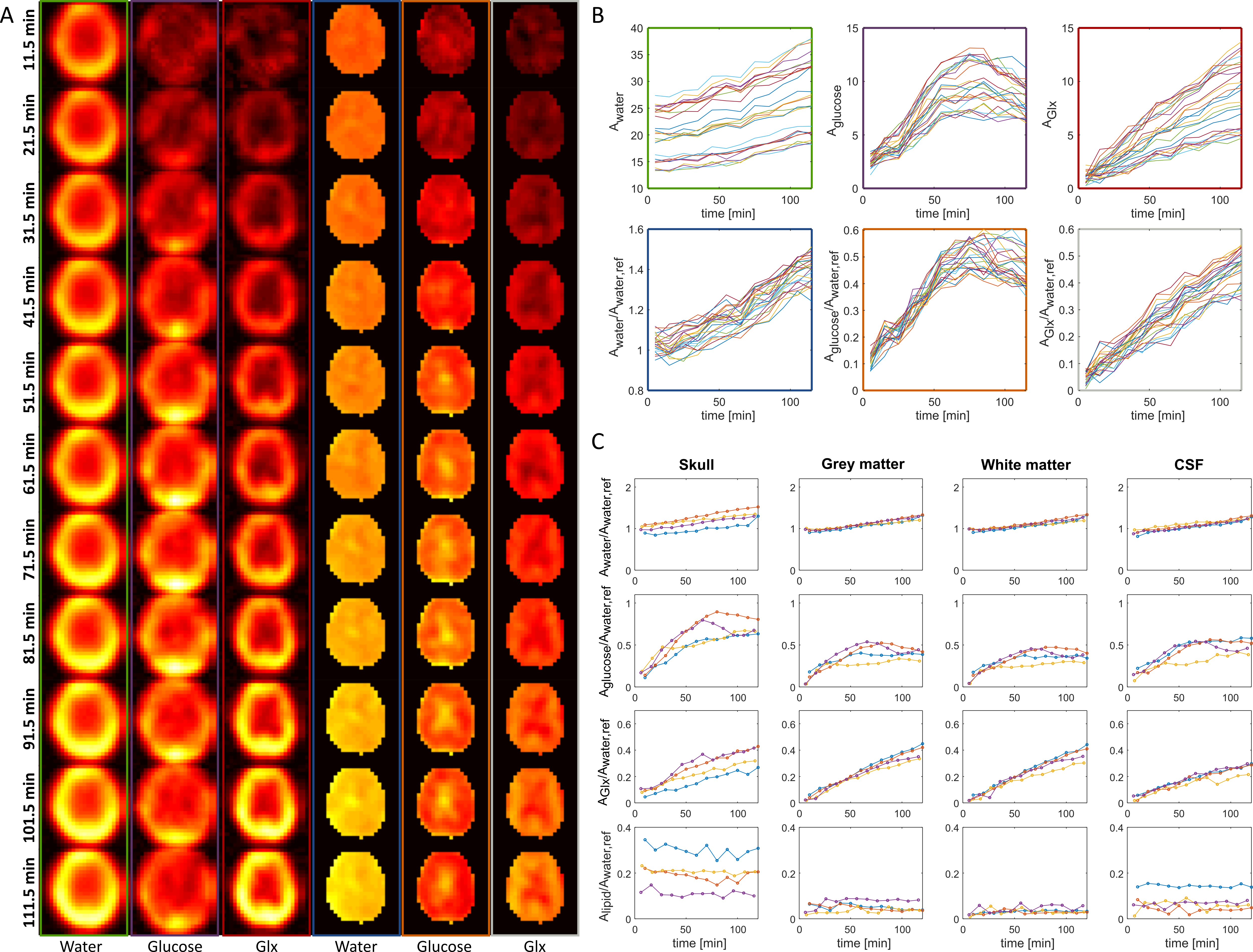
Fig. 5: 2H images for different times of one volunteer for
water, glucose and Glx with and without referencing to a baseline water
measurement (A). Zero-filling to twice the original resolution was
applied. In (B), signals for different
voxels are shown for water, glucose and Glx for the same volunteer. The colors
of the frame label the corresponding metabolic images in (A). (C) shows data
from four different volunteers (different colors) for water, glucose, Glx and
lipid/lactate for summed voxels over different regions of the head.
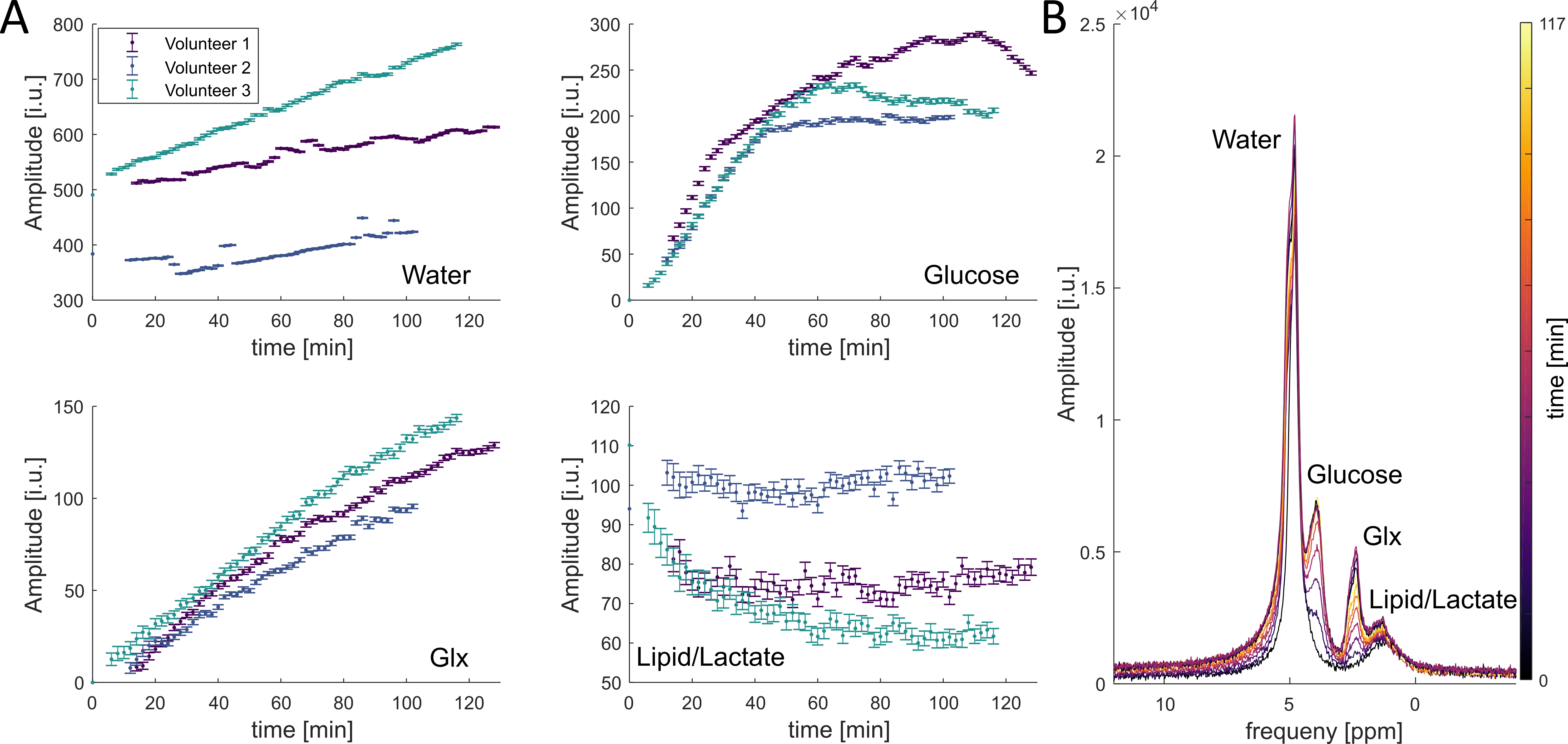
Fig. 2: The
results for three volunteers of a whole brain FID measurement after the oral
administration of deuterated glucose for four resonances (A). The temporal
resolution of the measurement is 2 minutes. In (B), several spectra from
volunteer 3 are shown at different time points. The time points are labeled with
different colors.