Qingping Chen1,2,3, Wieland A. Worthoff1, and N. Jon Shah1,2,4,5
1Institute of Neuroscience and Medicine 4, INM-4, Forschungszentrum Jülich, Germany, 2Department of Neurology, RWTH Aachen University, Aachen, Germany, 3Department of Biomedical Engineering, The University of Melbourne, Parkville, Australia, 4Institute of Neuroscience and Medicine 11, INM-11, JARA, Forschungszentrum Jülich, Germany, 5JARA - BRAIN - Translational Medicine, Aachen, Germany
1Institute of Neuroscience and Medicine 4, INM-4, Forschungszentrum Jülich, Germany, 2Department of Neurology, RWTH Aachen University, Aachen, Germany, 3Department of Biomedical Engineering, The University of Melbourne, Parkville, Australia, 4Institute of Neuroscience and Medicine 11, INM-11, JARA, Forschungszentrum Jülich, Germany, 5JARA - BRAIN - Translational Medicine, Aachen, Germany
The optimization of the triple-quantum-filtered sodium MRI sequence - enhanced SISTINA with FLORET improves the UTE image quality, while maintaining the multiple-quantum-filtered image quality and introducing randomness for potential application of compressed-sensing acceleration.
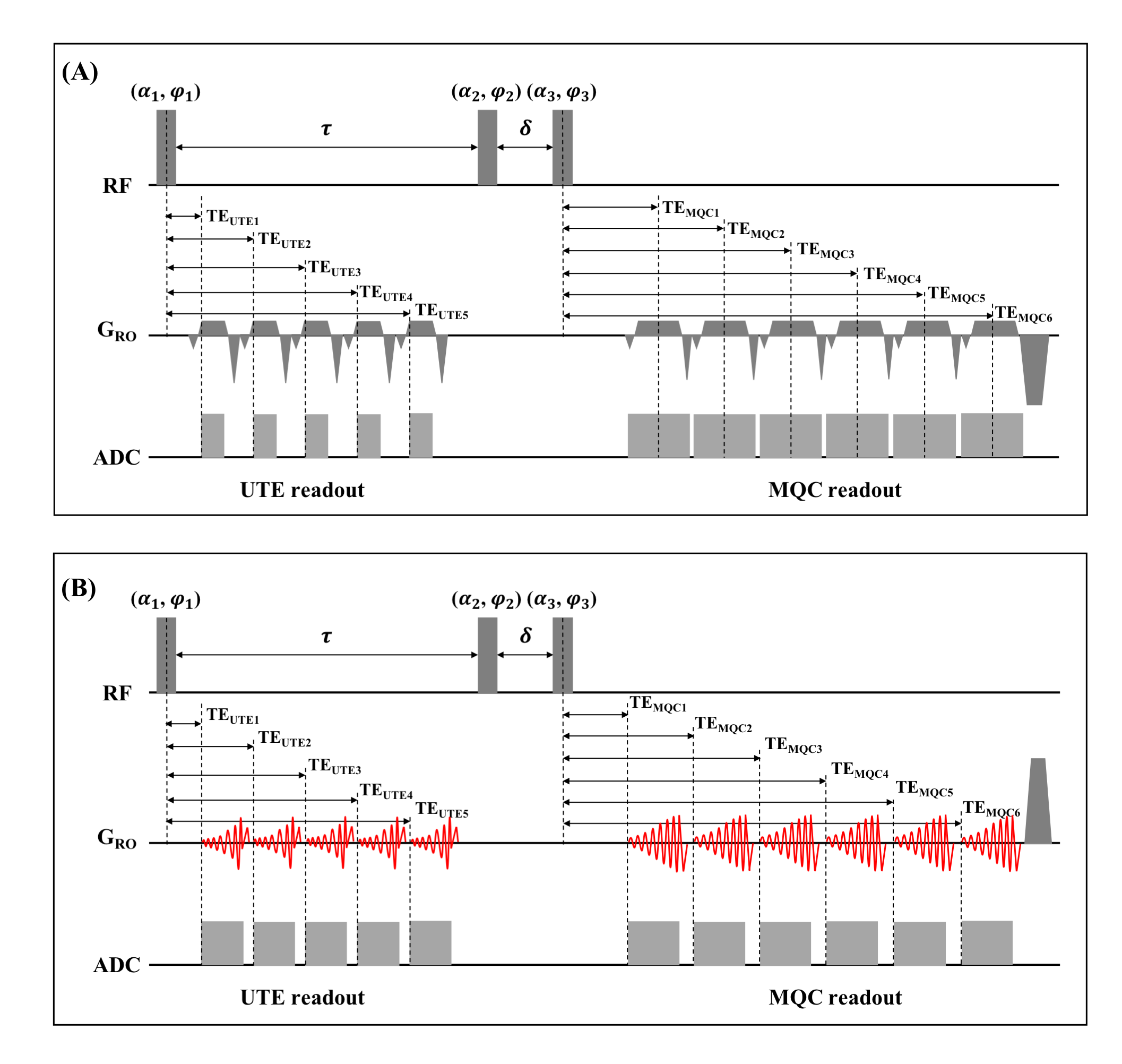
Figure 2: Sequence diagrams of (A) conventional enhanced SISTINA with DISCOBALL UTE and MGRE MQC readout, and (B) optimized enhanced SISTINA with FLORET UTE and MQC readouts. Three hard RF pulses with flip angle α1=α2=α3=90°, preparation time τ=10ms, evolution time δ=50us. 12-step phase cycling scheme is applied to separate SQ- and TQ-weighted signal. Rewinder gradients are applied immediately after every readout to avoid the effect of residual magnetization on high-order coherences. Spoilers are used to dephase the residual transversal magnetization after the last MQC readout.
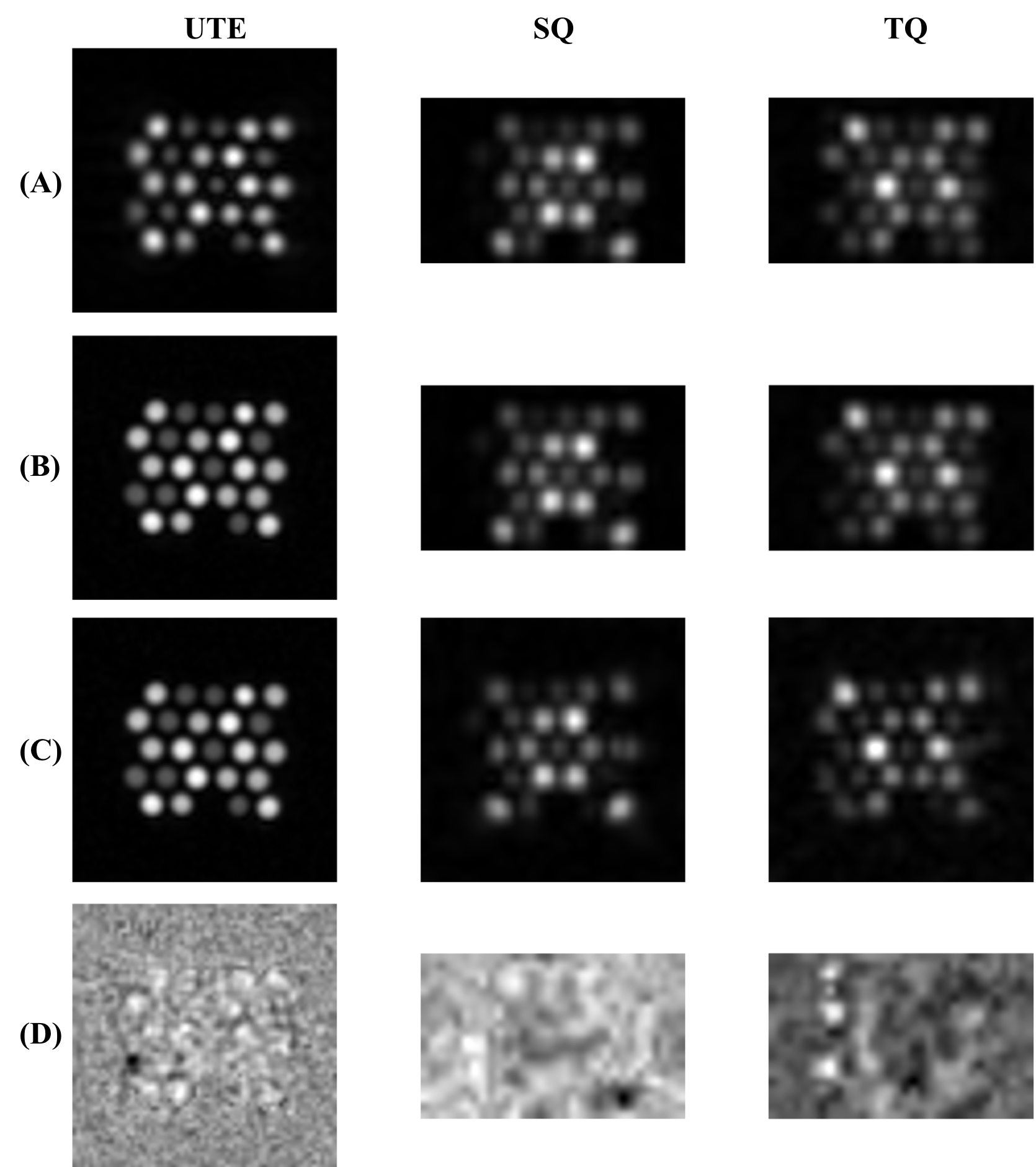
Figure 4: The first-echo UTE, SQ, and TQ images after B0 and B1 correction obtained from three measurements depicted in Table 1. (Left) UTE, (middle) SQ, and (right) TQ images of (A) conventional enhanced SISTINA, (B) enhanced SISTINA with FLORET UTE readouts and MGRE MQC readouts, and (C) optimized enhanced SISTINA with FLORET UTE and FLORET MQC readouts. (D) the image differences of UTE, SQ, and TQ images: (left) the UTE image difference between the second and third measurements; (middle) the SQ and (right) TQ image differences between the first and second measurements.