Yibo Zhao1,2, Rong Guo1,2, Yudu Li1,2, Keith R. Thulborn3, and Zhi-Pei Liang1,2
1Department of Electrical and Computer Engineering, University of Illinois, Urbana-Champaign, Urbana, IL, United States, 2Beckman Institute for Advanced Science and Technology, University of Illinois, Urbana-Champaign, Urbana, IL, United States, 3Center for Magnetic Resonance Research, University of Illinois at Chicago, Chicago, IL, United States
1Department of Electrical and Computer Engineering, University of Illinois, Urbana-Champaign, Urbana, IL, United States, 2Beckman Institute for Advanced Science and Technology, University of Illinois, Urbana-Champaign, Urbana, IL, United States, 3Center for Magnetic Resonance Research, University of Illinois at Chicago, Chicago, IL, United States
A
novel method is proposed for reconstruction of high-resolution sodium images
from noisy, limited k-space data. The proposed method has been validated using
both simulated and clinical data by comparison to conventional results.
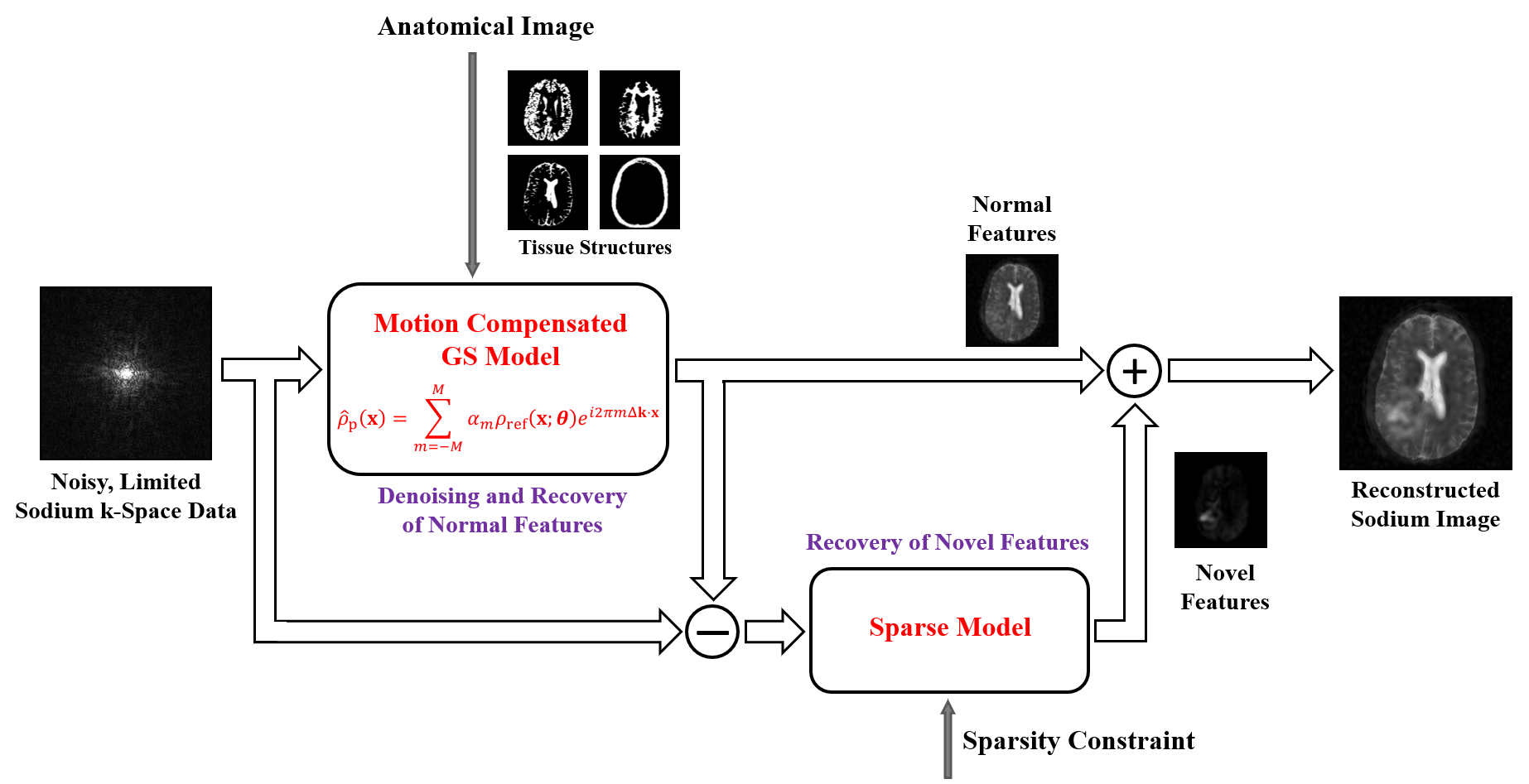
Figure 1. Illustration
of the proposed reconstruction method. Tissue-based structural information from
high-quality proton images is incorporated into the reconstruction of normal
image features using a motion-compensated GS model. Sodium-dependent novel
features (e.g., lesions) are reconstructed from the residues using a sparse
model.
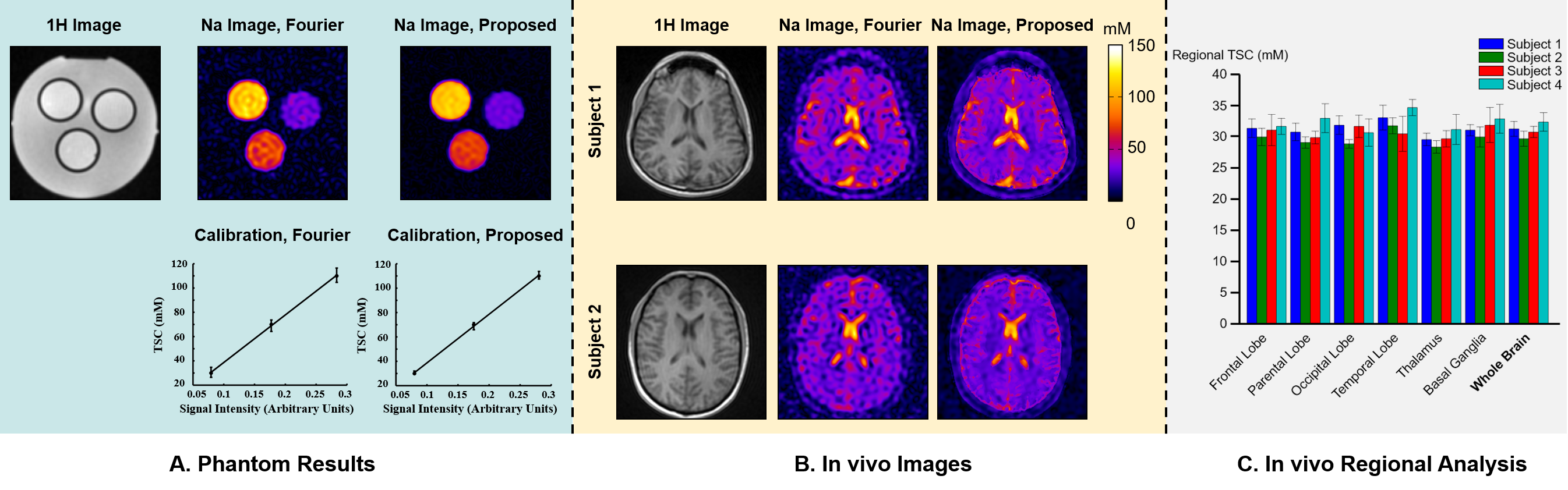
Figure 3. Phantom
and healthy subject results. (A) Phantom 1H reference image, 23Na FFT and proposed reconstruction, with corresponding calibration
curves. Note that both curves are linear,
but the FFT reconstruction had much larger standard deviation (5.3%) than
the proposed method (2.8%). (B) Images from two healthy subjects. Note the marked
improvement in image quality by the proposed method. (C) Regional TSC values obtained
using the proposed method, which were
consistent across different brain regions and subjects,
demonstrating the reproducibility of the proposed method.