Ali Caglar Özen1,2, Felix Spreter1, Timo Heidt3, Constantin von zur Mühlen3, and Michael Bock1
1Deptartment of Radiology, Medical Physics, University Medical Center Freiburg, University of Freiburg, Freiburg, Germany, 2German Consortium for Translational Cancer Research Partner Site Freiburg, German Cancer Research Center (DKFZ), Heidelberg, Germany, 3Department of Cardiology and Angiology I, UHZ, University Medical Center and Faculty of Medicine, University of Freiburg, Freiburg, Germany
1Deptartment of Radiology, Medical Physics, University Medical Center Freiburg, University of Freiburg, Freiburg, Germany, 2German Consortium for Translational Cancer Research Partner Site Freiburg, German Cancer Research Center (DKFZ), Heidelberg, Germany, 3Department of Cardiology and Angiology I, UHZ, University Medical Center and Faculty of Medicine, University of Freiburg, Freiburg, Germany
A detachable Tx array of as low as 5 independent loop coils can be used to generate a homogeneous B1+ field within the heart of an adult pig. In combination
with an optimized Rx coil array that conforms to the special geometry of a pig
supine position high SNR can be achieved.
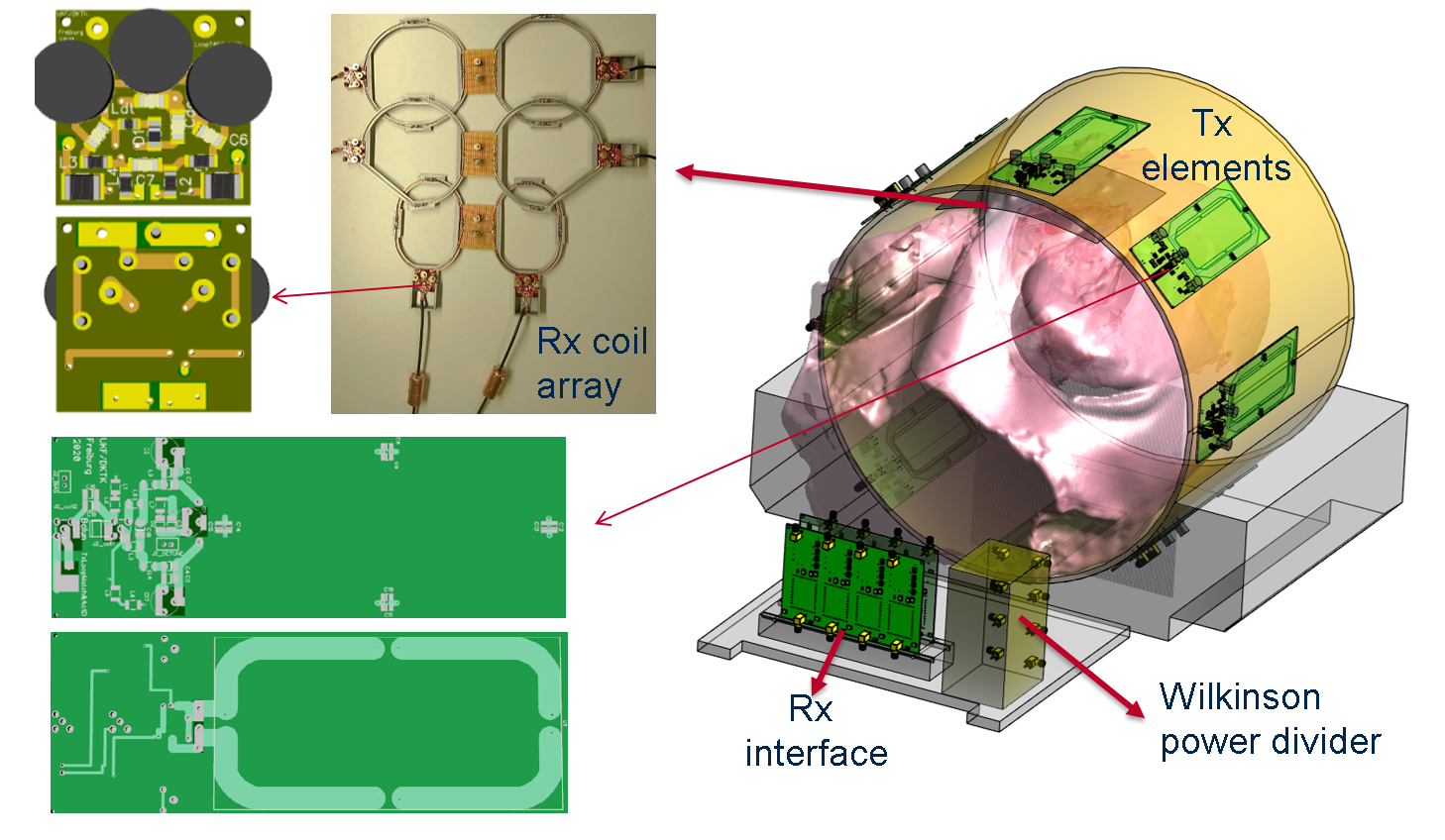
Fig.
1: Schematic and layout of the dedicated 19F MRI RF coil system. Printed
circuit boards of the Tx and Rx coil elements are shown on the left hand side. Sij curves, circuit diagrams and the component values
can be found in https://github.com/alibaz/F19Coils
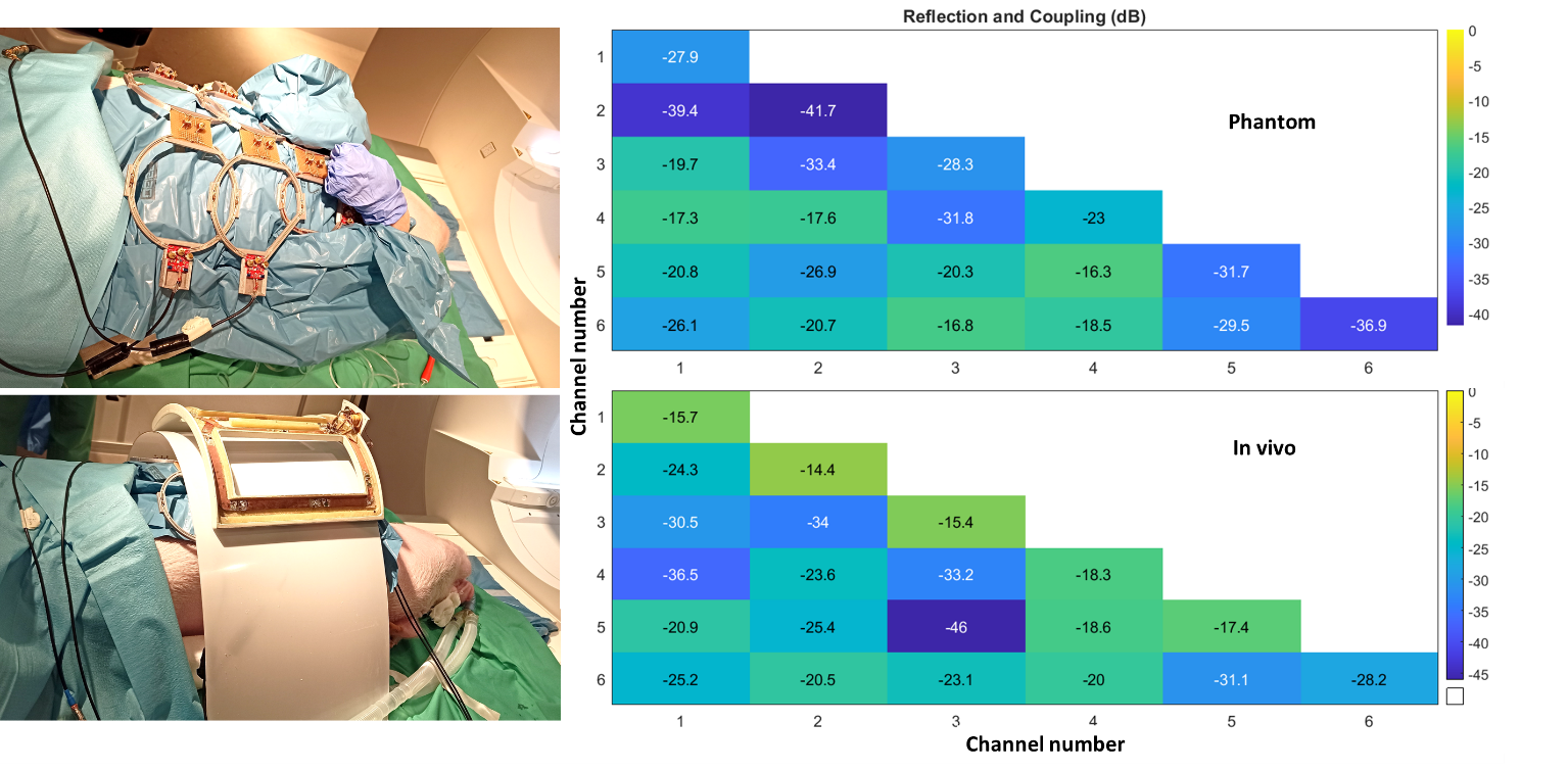
Fig.
4: S matrix of the 6 channel anterior part of the 19F MRI coil system measured
with phantom and in vivo loading. In the phantom Sii<-20dB and Sij<-16
dB, and in the animal an Sii<-14dB and Sij<-18 dB was
achieved.