Alan J. Wright1, Richard Mair1,2,3, Anastasia Tsyben1, and Kevin M. Brindle1,3,4
1CRUK Cambridge Institute, University of Cambridge, Cambridge, United Kingdom, 2Department of Clinical Neurosciences, University of Cambridge, Cambridge, United Kingdom, 3Cancer Research UK Major Centre-Cambridge, University of Cambridge, Cambridge, United Kingdom, 4Department of Biochemistry, University of Cambridge, Cambridge, United Kingdom
1CRUK Cambridge Institute, University of Cambridge, Cambridge, United Kingdom, 2Department of Clinical Neurosciences, University of Cambridge, Cambridge, United Kingdom, 3Cancer Research UK Major Centre-Cambridge, University of Cambridge, Cambridge, United Kingdom, 4Department of Biochemistry, University of Cambridge, Cambridge, United Kingdom
Tensor decomposition can be used for denoising magnetic resonance spectroscopic imaging (MRSI) data. A condition for selection of rank order is proposed that removes only noise from the reconstructed data and preserves signals from low amplitude resonances.
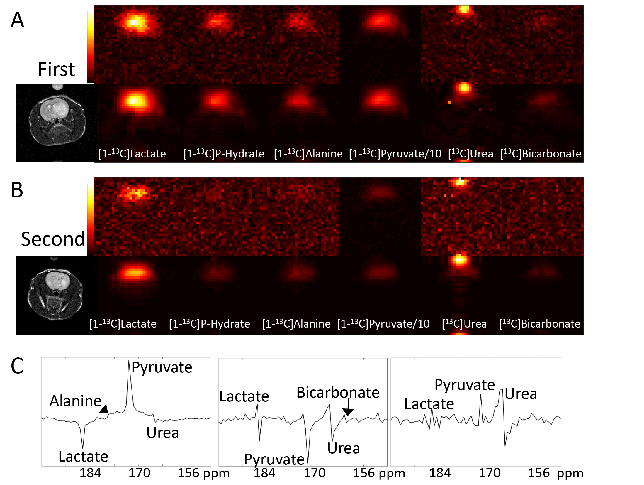
Figure 3. A) Metabolite
amplitudes of the first 13C MRSI dataset: top row - acquired data;
lower row - TRI de-noised data (core rank: 7x6x7). A T2-weighted
image shows the positions of the brain, tumour and a [13C]urea phantom.
Relative amplitudes are indicated by the colour bar, except for [1-13C]pyruvate,
which is decreased ten-fold. B) As for (A), but for the second dataset (core
rank 3x3x3). C) The first three fibers in the spectral dimension of the tensor
decomposition in (B), shown as spectra.
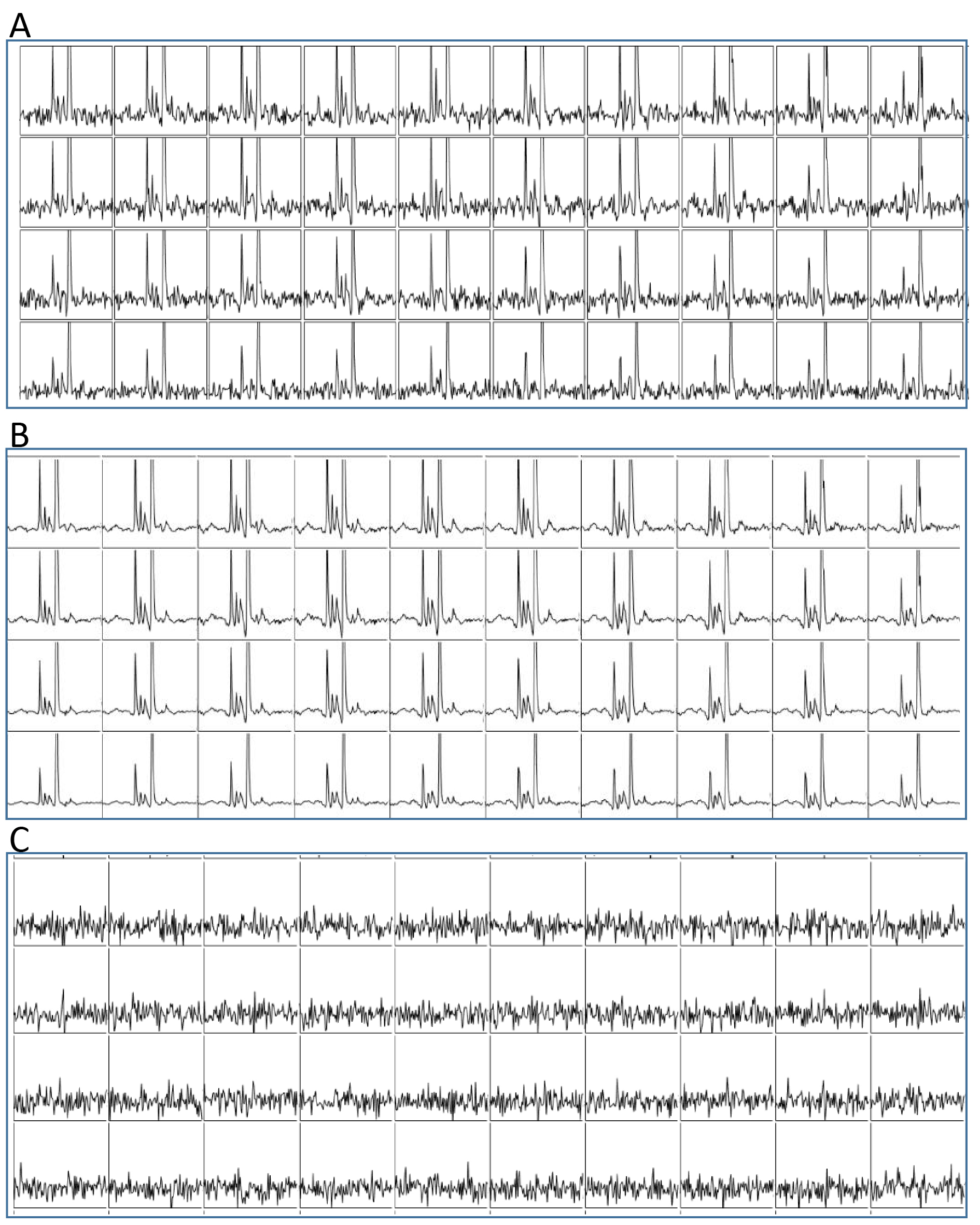
Figure 1. A) Carbon-13 MRSI spectra from the
head of a rat with an orthotopically implanted patient-derived glioblastoma
xenograft. Rows 7 to 10, columns 13 to 22 and chemical shifts 209 ppm to 141 ppm
are shown. Spectra were baseline corrected and line broadened to 25 Hz. B) The same data but reconstructed with a
core tensor size of 7x6x7 and processed similarly. C) The residual of the
acquired data minus the TRI data with no line broadening or baseline correction.