Parker John Bresnahan Jenkins1, Guangle Zhang1, Wei Zhu1, Xiao-Hong Zhu1, and Wei Chen1
1Center for Magnetic Resonance Research, University of Minnesota, Minneapolis, MN, United States
1Center for Magnetic Resonance Research, University of Minnesota, Minneapolis, MN, United States
A novel tri-frequency
surface coil based on traditional loop design utilizing active tuning circuitry
was developed and validated for 1H, 2H, and 17O
MR applications at 16.4T. It shows a potential for temporally efficient interleaved
metabolic imaging.
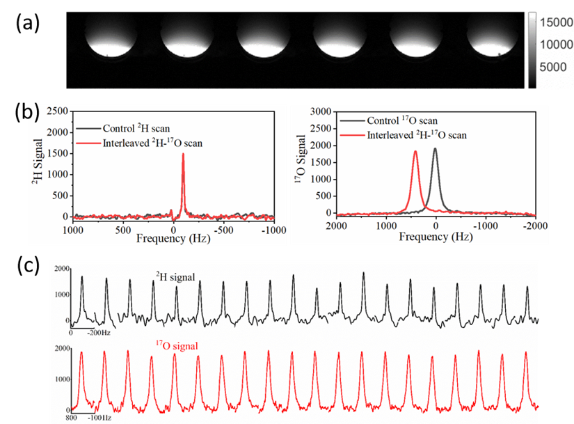
Figure 5. (a) 1H axial
images acquired with 2D GEMS sequence. (b) Left: the 2H
signal of Control scan and that of interleaved 2H- 17O
scans (nt=20). Right: the 17O signal of the Control scan and that
of interleaved 2H- 17O scans (nt=100). (c) Top: 20
deuterium spectra from interleaved 2H- 17O scans (nt=1×20);
Bottom: 20 oxygen-17 spectra from interleaved 2H- 17O
scans (nt=5×20).
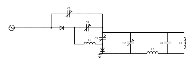
Figure 1. Schematic circuit diagram of the triple-tuned coil design. For 1H (no DC), modulate C2 and C5 to tune and match to 698MHz. For 2H (no
DC) tune and match C2 and C5 to 107MHz. For 17O, make sure setup for 2H is established, apply DC and modulate C3 and C4 to tune and match
to 94MHz.