Fabian Küppers1,2,3, Seong Dae Yun1, and N. Jon Shah1,2,4,5
1Institute of Medicine and Neuroscience 4, Forschungszentrum Juelich GmbH, Jülich, Germany, 2Institute of Medicine and Neuroscience 11, Forschungszentrum Juelich GmbH, Jülich, Germany, 3RWTH Aachen University, Aachen, Germany, 4Department of Neurology, RWTH Aachen University, Aachen, Germany, 5JARA - BRAIN - Translational Medicine, Aachen, Germany
1Institute of Medicine and Neuroscience 4, Forschungszentrum Juelich GmbH, Jülich, Germany, 2Institute of Medicine and Neuroscience 11, Forschungszentrum Juelich GmbH, Jülich, Germany, 3RWTH Aachen University, Aachen, Germany, 4Department of Neurology, RWTH Aachen University, Aachen, Germany, 5JARA - BRAIN - Translational Medicine, Aachen, Germany
Simultaneous
quantification
of T2 and T2* within
1 minute
using
improved
10-echo GESE-EPIK is
validated for phantoms
and in
vivo
with reference methods. Successful
WM/GM separation is
shown. Sequence acceleration
is
investigated
with
(t)SNR
analysis.
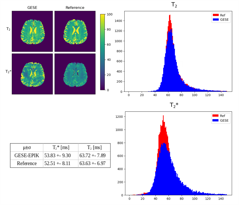
Figure 4: In
vivo
relaxation
quantification results. One exemplary slice for T2 and T2* maps from
GESE-EPIK and the reference methods is shown in the top left corner.
T2*
and T2
histograms from
all acquired slices
compare
each parameter distribution from GESE-EPIK with its reference method.
Mean values of each distribution with its standard deviation are
given.
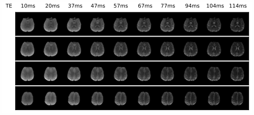
Figure 2: Reconstructed
images from 10-echo
GESE-EPIK for four exemplary slices of an in
vivo
data set. The
TE
for each echo is
given. The
signal
strength of later echoes is modulated for visualisation
purposes.