Florian Wiesinger1,2, Graeme McKinnon3, Sandeep Kaushik1, Ana Beatriz Solana1, Emil Ljungberg2, Mika Vogel1, Naoyuki Takei4, Rolf Schulte1, Carolin Pirkl1, Cristina Cozzini1, Laura Nuñez-Gonzalez5, Juan A. Hernandez Tamames5, and Mathias Engström6
1GE Healthcare, Munich, Germany, 2IoPPN, Department of Neuroimaging, King's College London, London, United Kingdom, 3GE Healthcare, Waukesha, WI, United States, 4GE Healthcare, Hino, Japan, 5Erasmus MC, Rotterdam, Netherlands, 6GE Healthcare, Stockholm, Sweden
1GE Healthcare, Munich, Germany, 2IoPPN, Department of Neuroimaging, King's College London, London, United Kingdom, 3GE Healthcare, Waukesha, WI, United States, 4GE Healthcare, Hino, Japan, 5Erasmus MC, Rotterdam, Netherlands, 6GE Healthcare, Stockholm, Sweden
3D Silent Parameter Mapping enables robust, high-resolution quantitative parameter mapping in the head. Beside silent neuroimaging the method also demonstrates unique potential for MR-only RTP in terms of synthetic CT conversion and automated organ-at-risk (OAR) delineation.
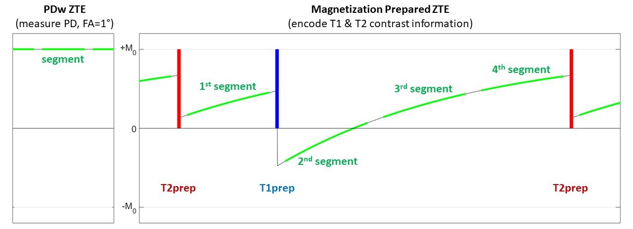
Figure 1: 3D Silent Parameter Mapping starts with a low FA~1° PD measurement (left) followed by a magnetization prepared segmented ZTE acquisition (FA=3°) to encode T1 and T2 contrast weighted information (right).
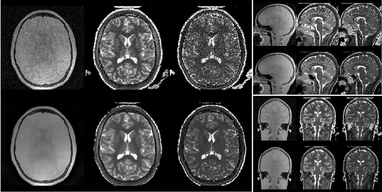
Figure 3: 3D Silent Parameter Mapping (FOV=19.2cm, res=1.2mm, BW=±31.25kHz, 1.5 averages, 6min05sec) at 1.5T showing PD (left), T1 (0…2s, middle) and T2 (0…1.6s,
right) parameter maps.