Jeffry R. Alger1,2,3,4, Jae Mo Park1, Junjie Ma1, Mahitha Roy1, Crystal Harrison1, James Ratnakar1, Albert Chen5, Galen Reed6, A. Dean Sherry1,7, Vlad Zaha1, and Craig R. Malloy1,8
1Advanced Imaging Research Center, University of Texas Southwestern Medical Center, Dallas, TX, United States, 2Neurology, University of California, Los Angeles, Los Angeles, CA, United States, 3NeuroSpectroScopics LLC, Sherman Oaks, CA, United States, 4Hura Imaging Inc, Los Angeles, CA, United States, 5GE Healthcare, Toronto, ON, Canada, 6GE Healthcare, Dallas, TX, United States, 7Chemistry, University of Texas at Dallas, Richardson, TX, United States, 8Cardiology, Veterans Affairs North Texas Healthcare System, Dallas, TX, United States
1Advanced Imaging Research Center, University of Texas Southwestern Medical Center, Dallas, TX, United States, 2Neurology, University of California, Los Angeles, Los Angeles, CA, United States, 3NeuroSpectroScopics LLC, Sherman Oaks, CA, United States, 4Hura Imaging Inc, Los Angeles, CA, United States, 5GE Healthcare, Toronto, ON, Canada, 6GE Healthcare, Dallas, TX, United States, 7Chemistry, University of Texas at Dallas, Richardson, TX, United States, 8Cardiology, Veterans Affairs North Texas Healthcare System, Dallas, TX, United States
Vascular dynamic simulations and preliminary human
investigations suggest a ‘patient-friendly’ infusion rate of 2.0 cm3/sec
is feasible for human heart metabolism studies that use hyperpolarized
pyruvate.
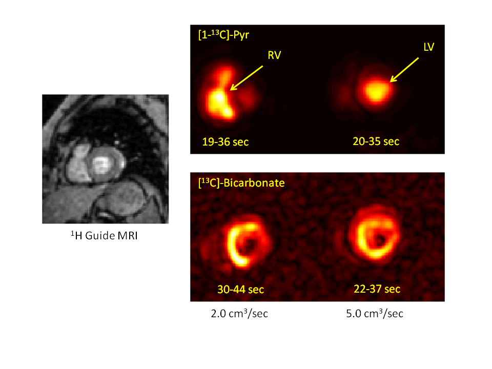
Figure 5: Dynamic MRSI of HP-[1-13C]Pyr and HP-[13C]-Bicarbonate
in human heart from infusion rates of 2.0 and 5.0 cm3/sec. Image
acquisition times following the start of infusion are shown below each image. The
slower infusion allows HP-[1-13C]Pyr visualization in the heart right
ventricle (RV) at the earliest feasible imaging time point, but for the faster
infusion HP-[1-13C]Pyr has mostly transited to the heart left ventricle by
this time.
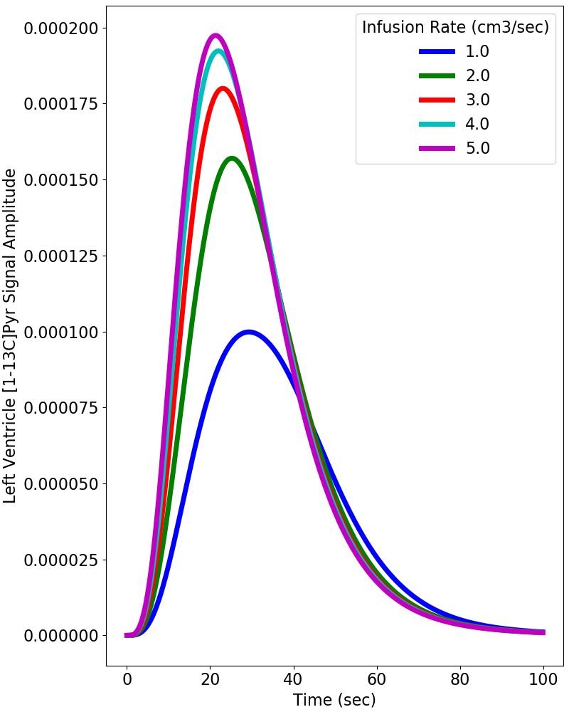
Figure 2: Dynamic simulations of the HP-[1-13C]Pyr
MR signal intensity (= concentration x polarization) in the heart left ventricle based on the model shown in Figure 1 using infusion rates
ranging from 1.0 to 5.0 cm3/sec.