Artem Mikheev1, Louisa Bokacheva1, Azadeh Sharafi1, Ravinder Regatte1, and Henry Rusinek1
1Department of Radiology, New York University School of Medicine, New York, NY, United States
1Department of Radiology, New York University School of Medicine, New York, NY, United States
A new elastic motion correction method was applied to the T1ρ-mapping
in 20 subjects with normal knees and early osteoarthritis. The T1ρ
voxel maps from motion-corrected images showed significantly lower variability in
articular cartilage compared to maps derived from uncorrected images.
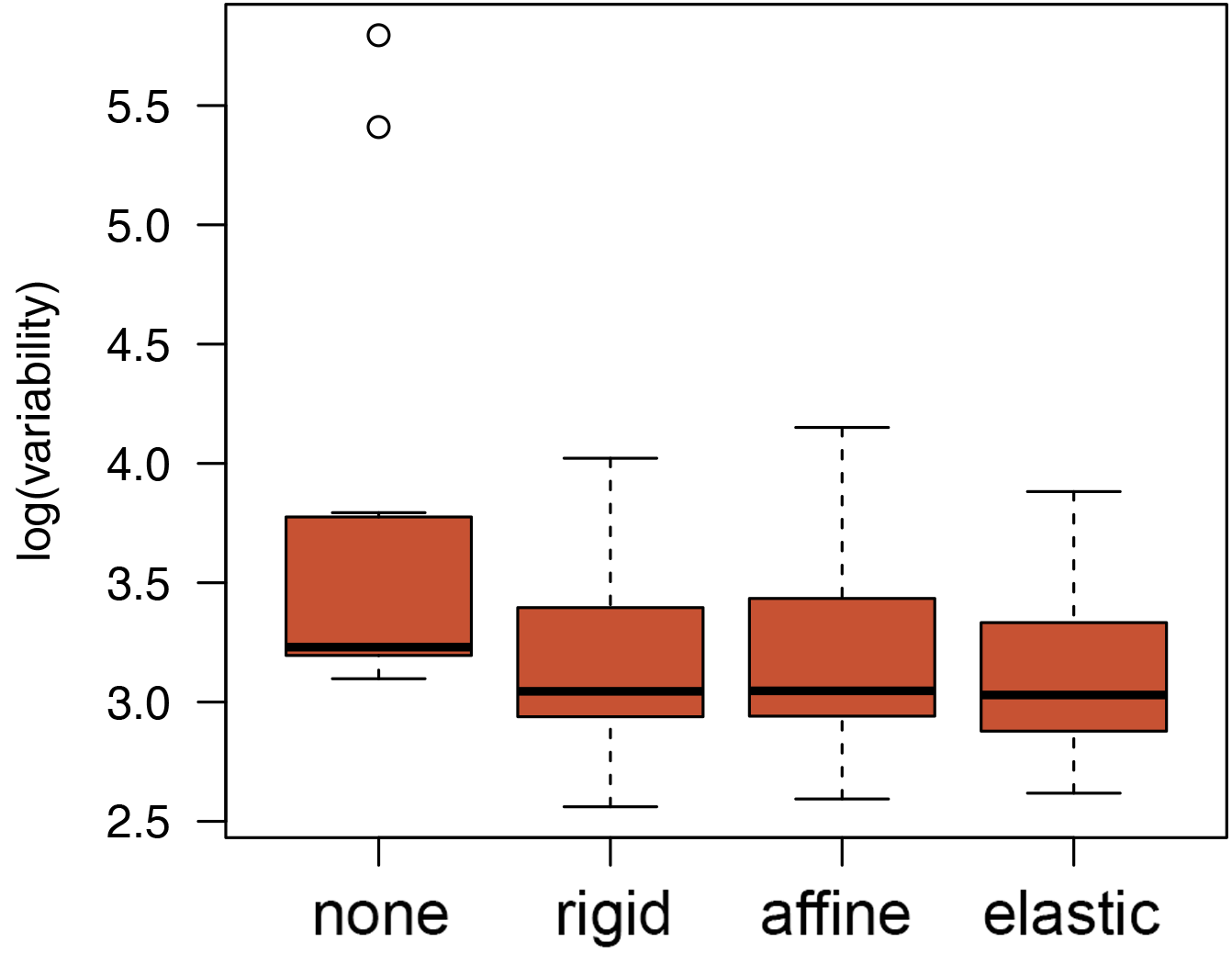
Figure 3: Box-and-whisker
plot of the T1ρ variability (standard deviation, log scale) in the
cartilage derived from images without motion correction (none) and images
corrected using three motion correction methods (rigid, affine, and elastic).
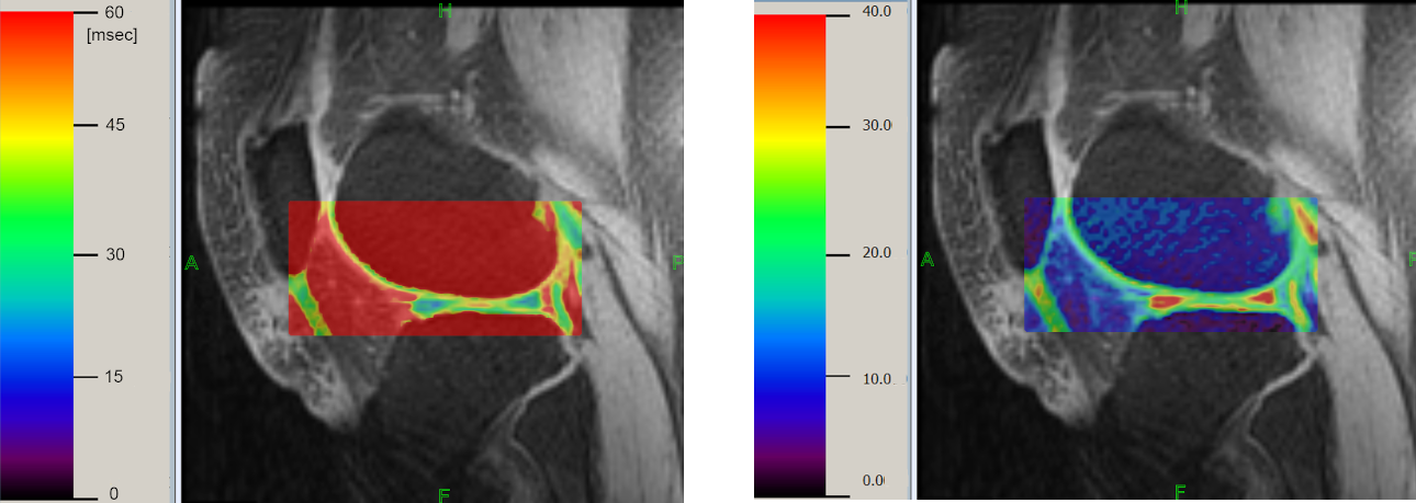
Figure 2: T1ρ (left,
color bar: ms) and R1ρ (right, color bar: s-1) maps of the cartilage derived after elastic registration.