Cheng-Chieh Cheng1, Jeffrey P. Guenette2, and Bruno Madore2
1Computer Science and Engineering, National Sun Yat-sen University, Kaohsiung, Taiwan, 2Radiology, Brigham and Women's Hospital, Harvard Medical School, Boston, MA, United States
1Computer Science and Engineering, National Sun Yat-sen University, Kaohsiung, Taiwan, 2Radiology, Brigham and Women's Hospital, Harvard Medical School, Boston, MA, United States
A quantitative and synthetic MRI method is being validated in its ability to deliver multiple quantitative and qualitative image contrasts, with 1-mm isotropic resolution and full brain coverage, in about 5.5 minute of scan time. Preliminary results are presented here in patients.
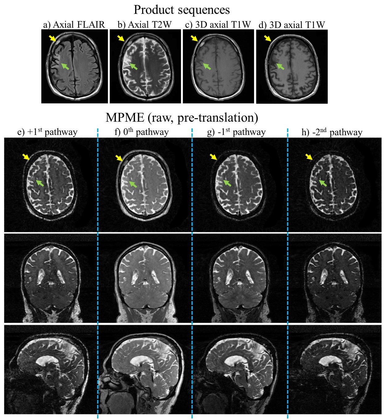
Fig. 2: Results from patient #2, using product sequences (a-d) and the present MPME sequence (e-h). MPME images are shown for all sampled pathways and a single echo time (out of three). These MPME images are not meant to be read directly, but contrast translation into traditional formats as shown in (a-d) will become possible only when the number of scanned patients is large enough for a neural network to be trained. An evolving infarct (green arrow) and a benign hemangioma (yellow arrow) are seen with varying degrees of conspicuity in the various contrasts and slices shown here.
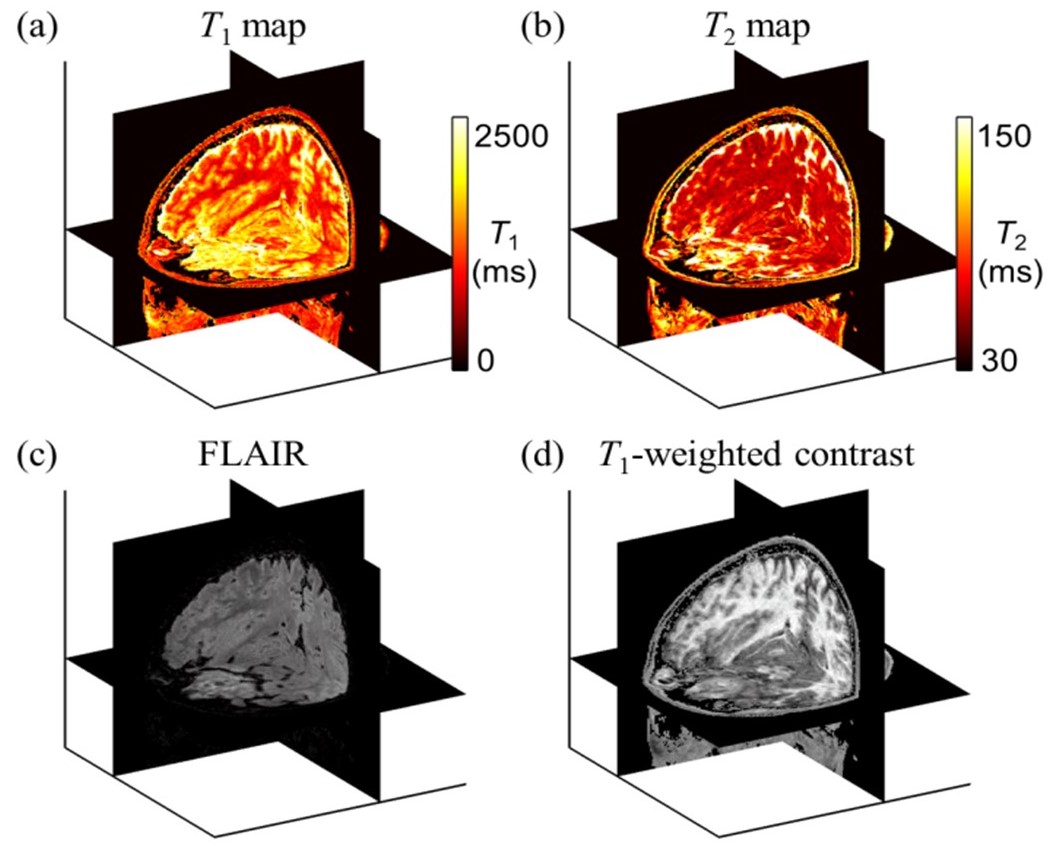
Fig. 1: Examples for four whole-head 3D contrasts generated through the neural translation of a single few-min MPME acquisition, from a prior healthy volunteer study: quantitative T1 (a) and T2 (b) maps in ms, and qualitative grayscale T2-FLAIR (c) and T1-weighted (d) contrasts. In addition to these four contrasts, MPME also readily generates susceptibility-weighted, proton-density weighted, T2-weighted and MPRAGE contrasts, along with quantitative flip angle maps.