Alexander M Barclay1, Michael C Langham1, and Felix W Wehrli1
1Department of Radiology, University of Pennsylvania, Philadelphia, PA, United States
1Department of Radiology, University of Pennsylvania, Philadelphia, PA, United States
An objective approach for background field inhomogeneity correction applied to field maps for susceptibility-based oximetry is proposed. Correction eliminates tissue phase. Venous oxygen saturation is measured using intravascular phase alone, reducing bias and increasing precision.
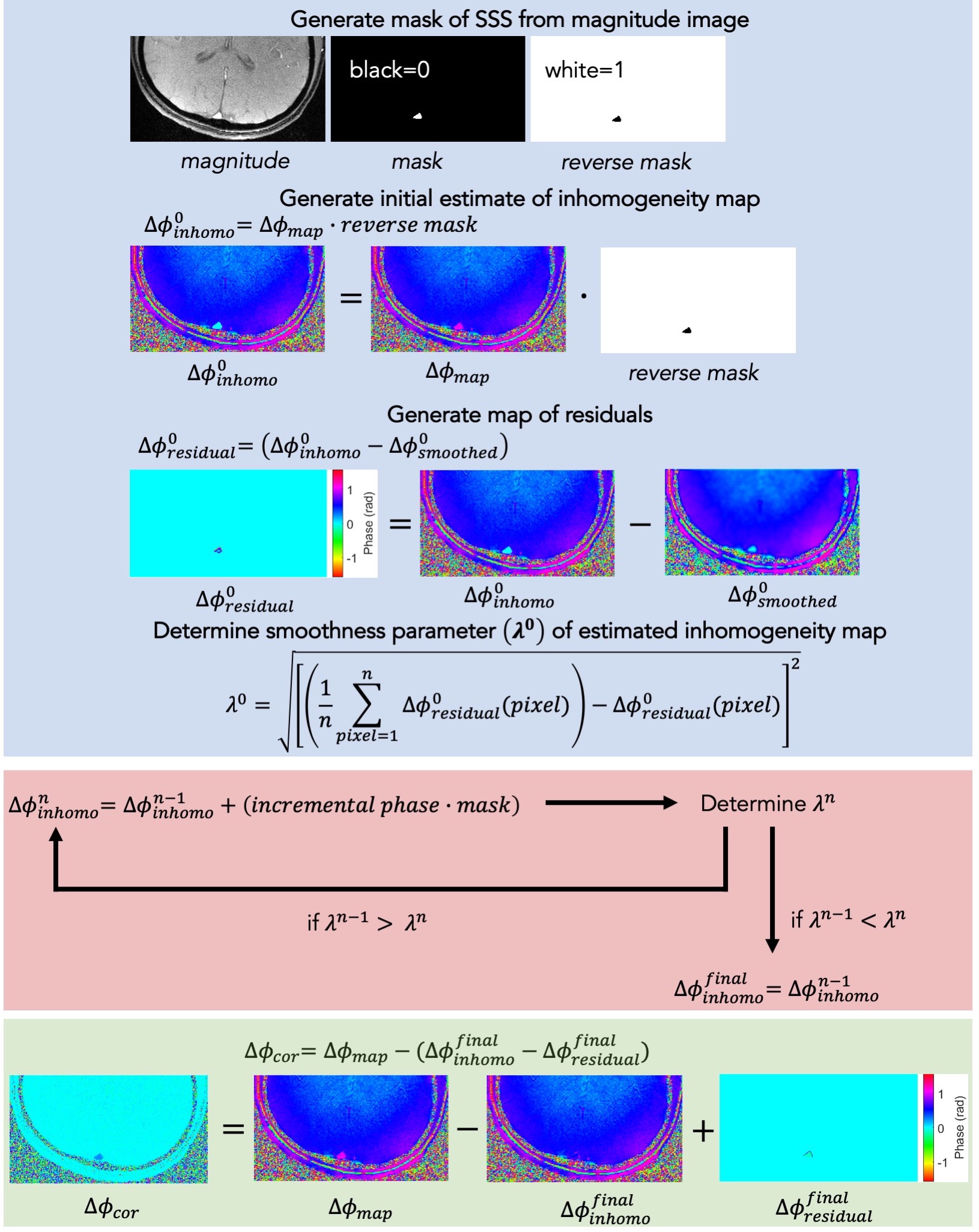
Figure 1: Smoothness-optimization algorithm for estimation of field inhomogeneity map. Blue panel: Preparation entails setting the initial estimate of the field inhomogeneity map, smoothing the estimate, generating a residual phase map, and calculating the smoothness parameter λ. Red panel: The background field inhomogeneity map is iteratively updated by addition of incremental phase and λ recalculated, until it is minimized. Green panel: The corrected field map is generated by subtraction of the final estimated inhomogeneity map, with final residual correction applied.
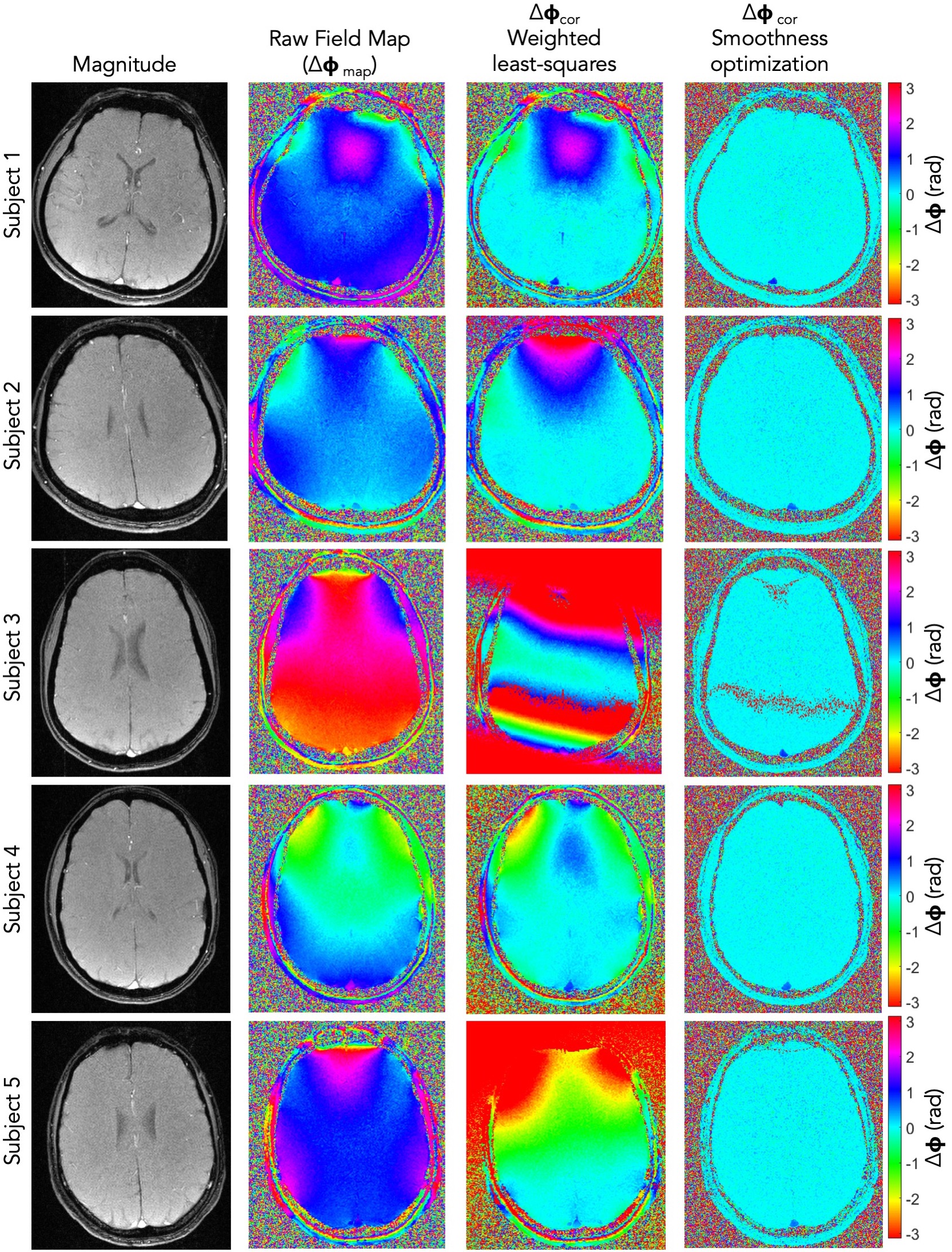
Figure 2: Field maps for five subjects. Data were acquired with the following parameters: FOV = 20 x 20 cm2; slice thickness = 5 mm; flip angle =12º; TR/ΔTE = 25/ 6.12 ms; bandwidth = 62.5 kHz; 200 phase-encodings; 200 readout points; reconstructed matrix = 400 x 400; voxel size = 1 x 1 x 5 mm3. Reconstruction performed according to Lee et al. (4). Column 1: magnitude image; column 2: raw field map; column 3: field-map corrected using weighted least-squares method; column 4: field-map corrected using smoothness optimization method.