Gunther Helms1,2, Lenka Vaculčiaková2, Kerrin J. Pine2, Harald E. Moeller3, and Nikolaus Weiskopf2,4
1Medical Radiation Physics, Clinical Sciences Lund, Lund University, Lund, Sweden, 2Neurophysics, Max Planck Institute for Human Cognitive and Brain Sciences, Leipzig, Germany, 3NMR Methods & Development Group, Max Planck Institute for Human Cognitive and Brain Sciences, Leipzig, Germany, 4Felix Bloch Institute for Solid State Physics, Leipzig University, Leipzig, Germany
1Medical Radiation Physics, Clinical Sciences Lund, Lund University, Lund, Sweden, 2Neurophysics, Max Planck Institute for Human Cognitive and Brain Sciences, Leipzig, Germany, 3NMR Methods & Development Group, Max Planck Institute for Human Cognitive and Brain Sciences, Leipzig, Germany, 4Felix Bloch Institute for Solid State Physics, Leipzig University, Leipzig, Germany
The multi-parameter mapping framework was used to estimate inhomogeneous MT (ihMT) effects in human brain at 3T in terms of fractional saturation of Mz. Experimental optimization was guided by an empirical signal equation. "ihMT saturation" was about 0.2% in WM at 100% SAR.
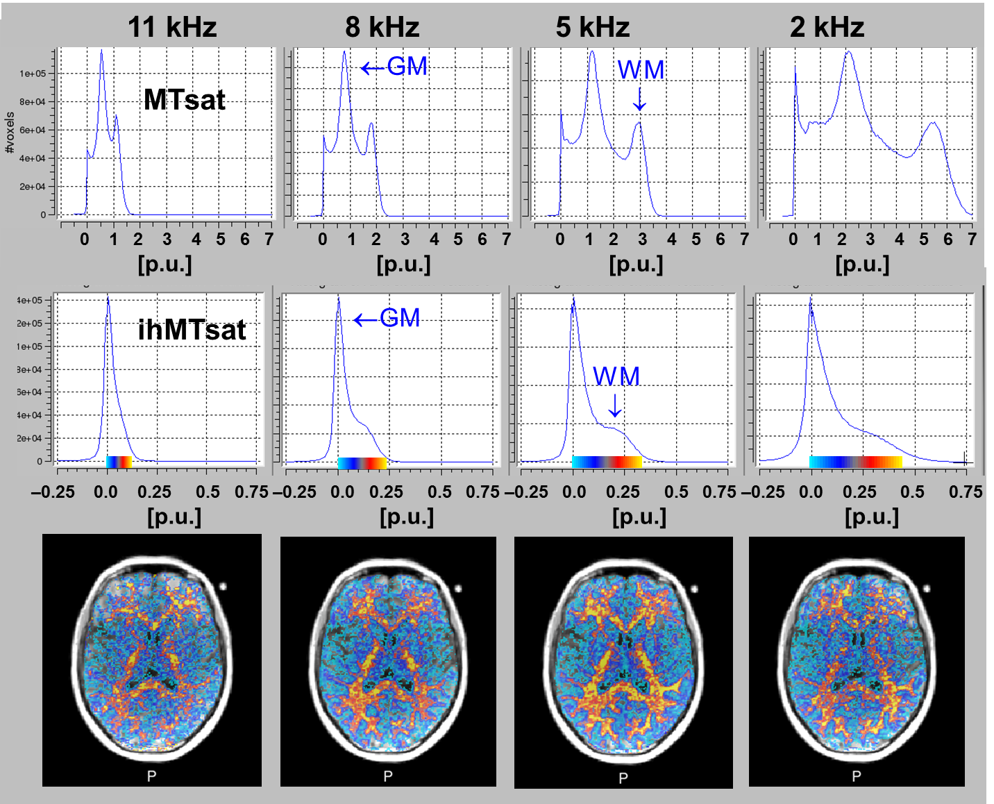
Figure 1: Offset dependence of MTsat and ihMTsat: Top: MTsat (--) increased
strongly towards small offsets. Middle: The right flank of the ihMTsat
histogram is assigned to WM. Its increases approximately parallel with WM MTsat.
Bottom: The ihMT maps at ±2kHz (right) showed a higher level of noise than at
5kHz, as judged from the overlay and histogram width This is explained by the
empirical signal equation (Eq. [1]) as high MTsat
(likely enhanced by direct saturation) compromises noise propagation.
The highest SNR in
the ihMTsat map was generally obtained around ±5kHz.

Figure 3: Increasing the SNR of ihMTsat maps.
Top row: ihMTsat calculated from averaged gradient echoes reveals regional differences in WM.
Middle row: These are corroborated by averaging two acquisitions with a 20-channel head coil. The histogram reveals a distinct WM mode around 0.2 p.u..
Bottom row: SNR is further improved by a 32-channel head coil, ±4kHz frequency offset and 10° readout. A distinct WM mode at slightly higher ihMTsat is seen despite minor motion artifacts.