Laurin Mordhorst1, Maria Morozova2,3, Sebastian Papazoglou1, Björn Fricke1, Jan Malte Oeschger1, Henriette Rusch3, Carsten Jäger2, Markus Morawski2,3, Nikolaus Weiskopf2,4, and Siawoosh Mohammadi1,2
1Institute of Systems Neuroscience, University Medical Center Hamburg-Eppendorf, Hamburg, Germany, 2Department of Neurophysics, Max Planck Institute for Human Cognitive and Brain Sciences, Leipzig, Germany, 3Paul Flechsig Institute of Brain Research, University of Leipzig, Leipzig, Germany, 4Felix Bloch Institute for Solid State Physics, Faculty of Physics and Earth Sciences, Leipzig, Germany
1Institute of Systems Neuroscience, University Medical Center Hamburg-Eppendorf, Hamburg, Germany, 2Department of Neurophysics, Max Planck Institute for Human Cognitive and Brain Sciences, Leipzig, Germany, 3Paul Flechsig Institute of Brain Research, University of Leipzig, Leipzig, Germany, 4Felix Bloch Institute for Solid State Physics, Faculty of Physics and Earth Sciences, Leipzig, Germany
We employed automated estimation of the effective axon radius ($$$r_{eff}$$$) on large-scale light microscopy data (>= 1mm²), showed that reliable estimation of $$$r_{eff}$$$ requires a large field-of-view and verified the potential of $$$r_{eff}$$$ to capture anatomical variation.
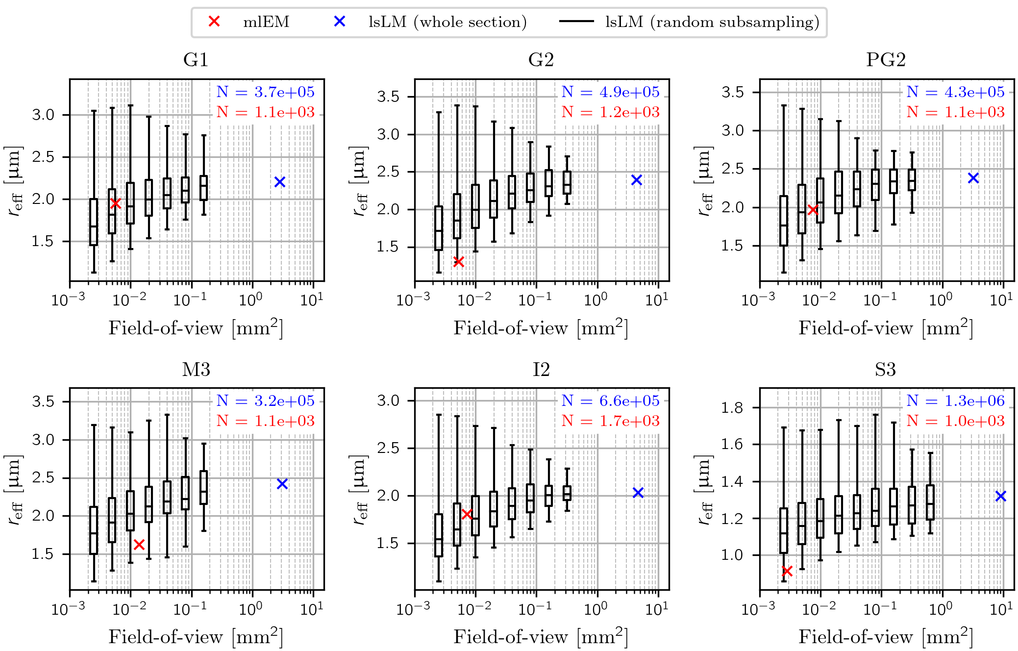
Figure 3: Influence of the FoV on $$$r_{\text{eff}}$$$. For six regions of the corpus callosum, box plots (horizontal lines in boxes denote median values; boxes include central 50 % of the values; whiskers include central 95 % of the values) of $$$r_{\text{eff}}$$$ for random lsLM subsections (black) are shown for varying FoV. The corresponding $$$r_{\text{eff}}$$$ of the whole lsLM sections (blue) and their consecutively cut mlEM subsections (red) are marked. The number of axons ranged from 1.0*10³ to 1.7*10³ in mlEM and 3.2*10⁵ to 1.3*10⁶ in lsLM.
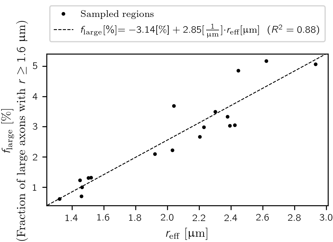
Figure 4: Relationship between $$$r_{\text{eff}}$$$ and the fraction of large axons. Point markers denote estimations of $$$r_{\text{eff}}$$$ from large-scale light microscopy (lsLM) sections of a human corpus callosum. The linear regression line ($$$f_{\text{large}}$$$ [%] = -3.14 [%] + 2.85 * [1/µm] * $$$r_{\text{eff}}$$$ [µm], R² = 0.88) is plotted as a dashed line. The number of identified axons per region ranged from 2.5*10⁴ to 1.3*10⁶ (on average: 4*10⁵).