Paul Han1, Thibault Marin1, Yanis Djebra1,2, Georges El Fakhri1, Jinsong Ouyang1, and Chao Ma1
1Gordon Center for Medical Imaging, Department of Radiology, Massachusetts General Hospital and Harvard Medical School, Boston, MA, United States, 2LTCI, Télécom Paris, Institut Polytechnique de Paris, Paris, France
1Gordon Center for Medical Imaging, Department of Radiology, Massachusetts General Hospital and Harvard Medical School, Boston, MA, United States, 2LTCI, Télécom Paris, Institut Polytechnique de Paris, Paris, France
This work presents a subspace-based fast MR method for free-breathing multi-delay ASL imaging of the kidney. The feasibility of the proposed method is shown using in vivo data obtained from a healthy volunteer on a 3T MR scanner.
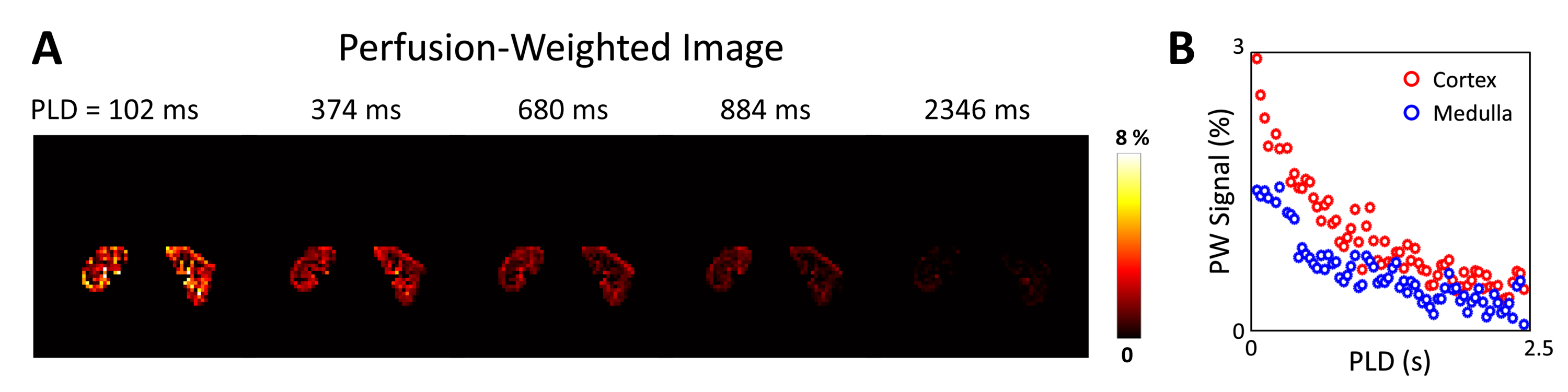
Figure 5. Perfusion-Weighted Image and Signal of
Kidney. A: Perfusion-weighted
images of the kidney over different PLD times. B: Mean perfusion-weighted
signal of the kidney cortex (red circle) and medulla (blue circle) over
different PLD times.
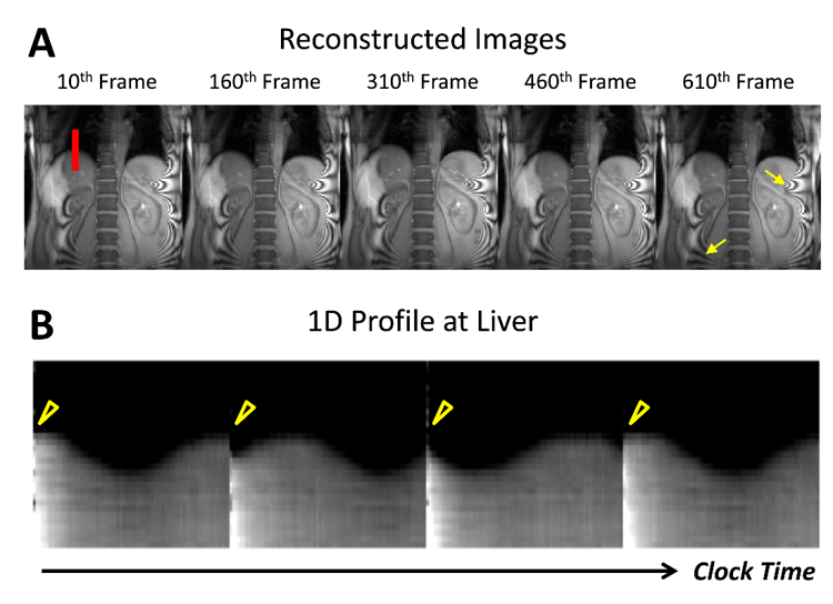
Figure 3. Results of reconstructed images over time. A: Reconstructed images at the 10th, 160th, 310th,
460th, and 610th frames. Yellow arrows indicate Moiré artifacts physically existing in the image due to the acquisition settings, which
are not originating due to subspace-based reconstruction. B: Evolution of 1D profile at the center of liver (red line in A) over clock time (frame rate 34 ms, frames 301 to 600). Notice the
change in liver position across time reflecting respiratory motion and the discontinuity in the liver position across time (yellow arrowhead) due to pCASL pulse.