Hui Liu1, Gaofeng Shi1, Qinglei Shi2, Weishuai Wang3, Jiangyang Pan1, and Yang Li1
1Fourth Hospital of Hebei Medical University, shijiazhuang, China, 2MR Scientific Marketing, Siemens Healthcare, beijing, China, 3CS, Services,, Siemens Healthcare, jinan, China
1Fourth Hospital of Hebei Medical University, shijiazhuang, China, 2MR Scientific Marketing, Siemens Healthcare, beijing, China, 3CS, Services,, Siemens Healthcare, jinan, China
The
sequence that continuously acquired Golden-angle RAdial Sparse Parallel
acquisition employing compressed sensing reconstruction (“GRASP”) can acquire
high spatial and high temporal resolution as well as motion robustness to DCE
MRI in liver imaging. However, there are still some artifacts in abdominal
imaging, especially in the early arterial phase. In this study, we proposed an
optimization scheme which can significantly improve the image quality both
in plain and all enhanced phases, which
may have important value in the study of abdominal disease using GRASP based DCE
in future.
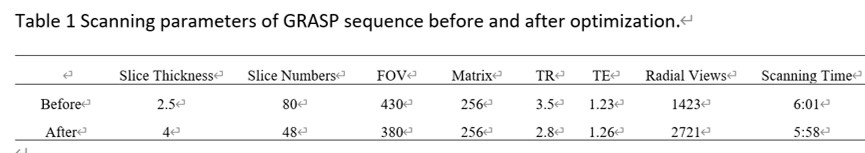
Table 1 Scanning parameters of GRASP
sequence before and after optimization.
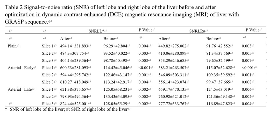
Table
2 Signal-to-noise ratio (SNR) of left lobe and right lobe of the liver before
and after optimization in dynamic contrast-enhanced (DCE) magnetic resonance
imaging (MRI) of liver with GRASP sequence.