Chau Vu1, Jian Shen1, Matthew Borzage2, Soyoung Choi3, and John Wood4
1Biomedical Engineering, University of Southern California, Los Angeles, CA, United States, 2Fetal and Neonatal Institute, Children's Hospital Los Angeles, Los Angeles, CA, United States, 3Neuroscience Graduate Program, University of Southern California, Los Angeles, CA, United States, 4Pediatrics and Radiology, Children's Hospital Los Angeles, Los Angeles, CA, United States
1Biomedical Engineering, University of Southern California, Los Angeles, CA, United States, 2Fetal and Neonatal Institute, Children's Hospital Los Angeles, Los Angeles, CA, United States, 3Neuroscience Graduate Program, University of Southern California, Los Angeles, CA, United States, 4Pediatrics and Radiology, Children's Hospital Los Angeles, Los Angeles, CA, United States
Using sinusoidally modulated CO2 stimulus which has previously been used to measure cerebrovascular reserve, this study derived a method to measure cerebral blood flow (CBF), cerebral blood volume (CBV) and transit time (TT) without the use of exogenous contrast.
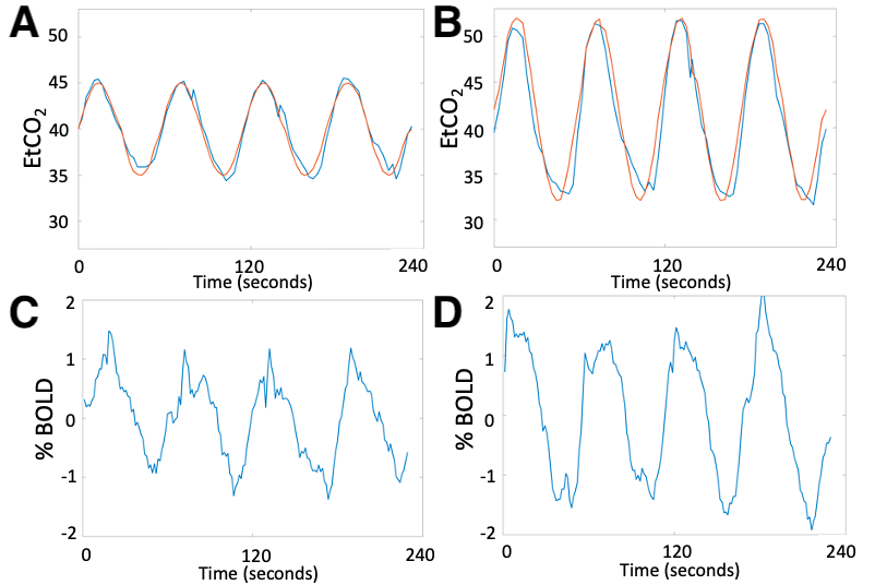
Figure 1. CO2 respiratory challenge with sinusoidally modulated stimulus in Subject 1. (A) Expected (red) and measured (blue) end-tidal CO2 with 5mmHg amplitude in paradigm #1. (B) Expected and measured end-tidal CO2 with 10mmHg amplitude in paradigm #2. (C) Whole brain BOLD time series in paradigm #1. (D) Whole brain BOLD time series in paradigm #2.
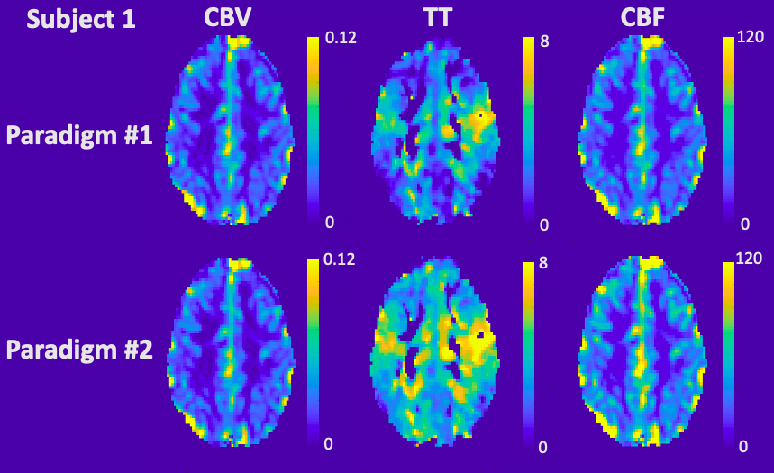
Figure 2. Perfusion maps derived from sinusoidal CO2 challenge in Subject 1. CBV, TT and CBF maps in paradigm #1 (top row) and paradigm #2 (bottom row).