Hongru Jia1, Chang Liu1, Weiqiang Dou2, Jing Ye1, and Xianfu Luo1
1Northern Jiangsu People’s Hospital, Yangzhou, China., Yangzhou, China, 2GE Healthcare,MR Research China, Beijing, China., Beijing, China
1Northern Jiangsu People’s Hospital, Yangzhou, China., Yangzhou, China, 2GE Healthcare,MR Research China, Beijing, China., Beijing, China
Multi-TE UTE imaging has been demonstrated to accurate measure R2* value at severe iron accumulation. It might be useful for clinical diagnosis of grading liver iron overload to guide iron chelation therapy.
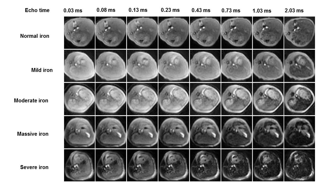
Figure 1. Representative multi-echo UTE liver images at varied degrees of iron overload in a rabbit model. With the iron deposition degree increasing, UTE liver signal decreased gradually. With the increase of echo times, UTE liver signal decreased gradually.
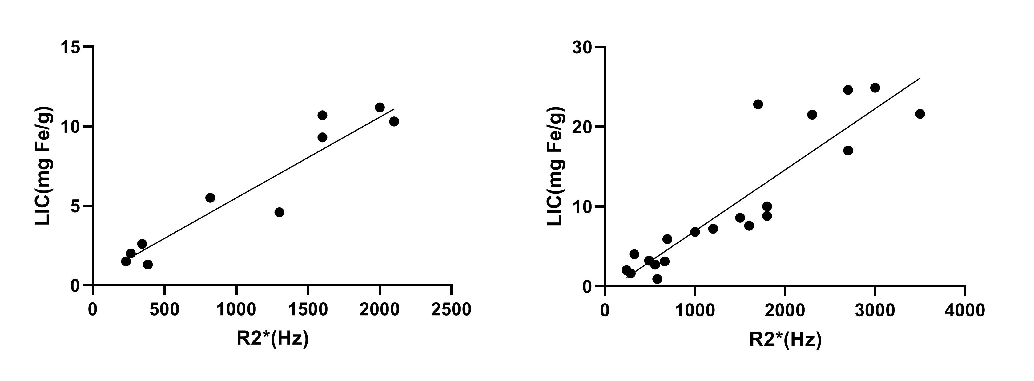
Figure 2. Correlation between hepatic R2* and liver iron content (LIC) in the normal fat liver group (A) and fatty liver group(B). The correlation coefficients were 0.911 and 0.811, respectively.