Mark Zamskiy1, Dominik Weidlich1, Kilian Weiss2, Marcus Makowski1, Rickmer Braren1, and Dimitrios Karampinos1
1Department of Diagnostic and Interventional Radiology, School of Medicine, Technical University of Munich, Munich, Germany, 2Philips Healthcare, Hamburg, Germany
1Department of Diagnostic and Interventional Radiology, School of Medicine, Technical University of Munich, Munich, Germany, 2Philips Healthcare, Hamburg, Germany
The present work introduced a novel T2-prepared radial SoS gradient echo sequence and demonstrated its applications: fat suppressed volumetric T2-weighted imaging in the combination with a 2-echo DIXON readout and the possibility for 3D isotropic resolution T2 mapping in the liver.
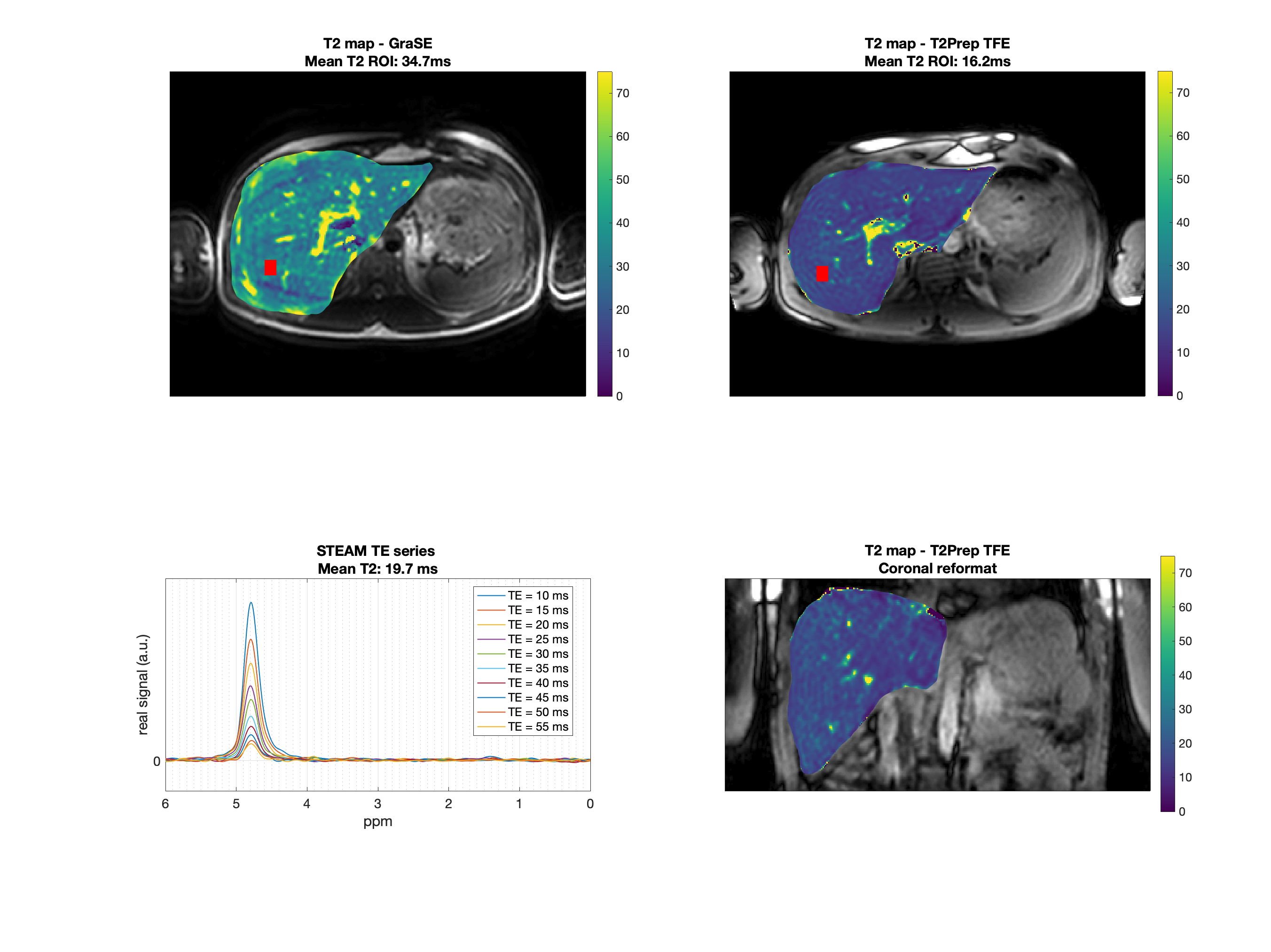
Fig.4. First row: Axial T2 maps obtained from GraSE and T2-prepared TFE. The red box represents the MRS voxel. Second row: MRS and the T2-prepared TFE coronal reformatted T2 map. Mean ROI value represents the mean value within the MRS acquisition voxel. T2 values from the imaging sequences deviate by 76 % (GraSE) and 18% (T2-prepared TFE) from MRS. Artifacts due to motion are visible in the GraSE but not in the T2-prepared TFE sequence.
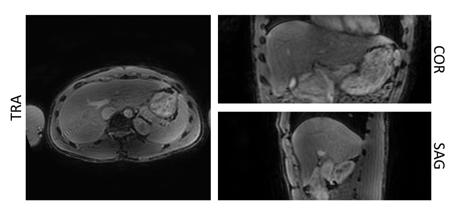
Fig.3. Separated T2-weighted water images obtained from the T2-prepared TFE acquisition with DIXON readout for with an effective echo time of the T2-preparation of 15ms. The fat signal is effectively suppressed, and the resulting isotropic resolution T2-weighted image allows reformatting in all 3 planes.