Hannah Grace Williams1,2, Caroline Hoad1,3, Neele Dellschaft1,3, Christabella Ng3,4, Alan Smyth3,4, Giles Major2,3, and Penny Gowland1,3
1Sir Peter Mansfield Imaging Centre, University of Nottingham, Nottingham, United Kingdom, 2Nottingham Digestive Diseases Centre, University of Nottingham, Nottingham, United Kingdom, 3National Institute for Health Research (NIHR) Nottingham Biomedical Research Centre, Nottingham University Hospitals NHS Trust and the University of Nottingham, Nottingham, United Kingdom, 4Division of Child Health, Obstetrics and Gynaecology, University of Nottingham, Nottingham, United Kingdom
1Sir Peter Mansfield Imaging Centre, University of Nottingham, Nottingham, United Kingdom, 2Nottingham Digestive Diseases Centre, University of Nottingham, Nottingham, United Kingdom, 3National Institute for Health Research (NIHR) Nottingham Biomedical Research Centre, Nottingham University Hospitals NHS Trust and the University of Nottingham, Nottingham, United Kingdom, 4Division of Child Health, Obstetrics and Gynaecology, University of Nottingham, Nottingham, United Kingdom
We developed
an MRI protocol to study the initial process of faeces formation in the colon. This
will provide new insights into colonic function, the process of faecalization
throughout the gastrointestinal tract and growth and proliferation of the
microbiota.
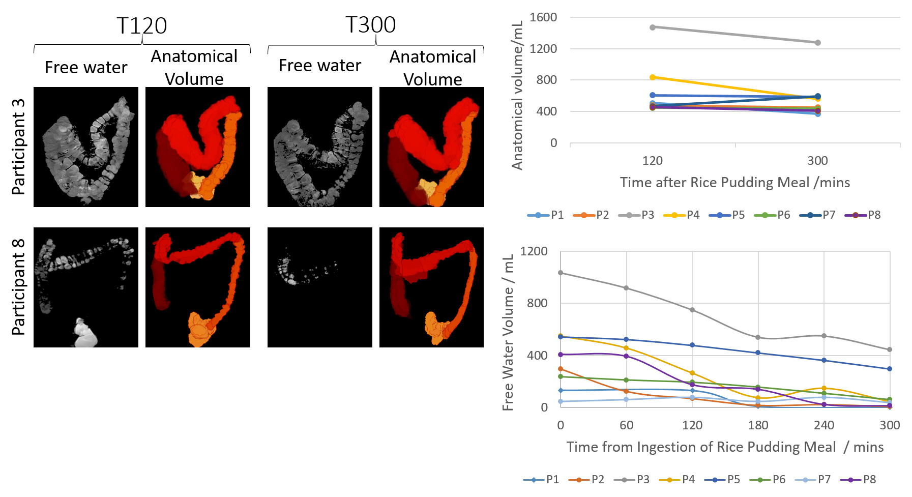
Figure 3: Free
water image and 3D rendering of colonic volume taken from participants 3 and 8
at 120 and 300 after the rice pudding meal. Plots show the colonic volume
and water content measurements for all participants.
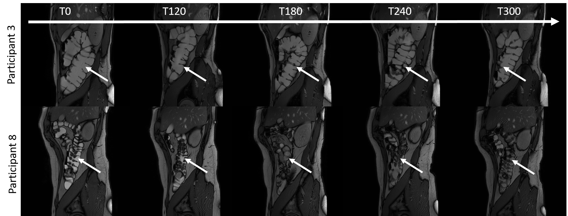
Figure 2: High
resolution images taken of the colon in participants 3 and 8. The colon is
indicated by the small white arrows.