hang qu1, wenjuan ba1, weiqiang Dou2, and wei wang1
1The Affiliated Hospital of Yangzhou University, yangzhou, China, 2GE Healthcare, MR Research China, Beijing, Beijing, China
1The Affiliated Hospital of Yangzhou University, yangzhou, China, 2GE Healthcare, MR Research China, Beijing, Beijing, China
在这项研究中,我们发现T2DM患者皮质区域的CBF缺陷与先前的研究一致。GIS1的灌注特征在双侧枕上回,楔形叶和顶顶回中表现出不同的灌注方式,并且其特征值与空腹血糖呈正相关。这些区域主要位于钙化裂隙周围,包括主要和次要视觉皮层,横纹皮层,超横纹皮层等。GIS2灌注的特征区域是双侧前扣带回,尾状核头,内囊前肢,壳状核等,这可能是由于穿孔动脉更深和对血管病理的敏感性更高(例如局部缺血或灌注不足) )[3]。
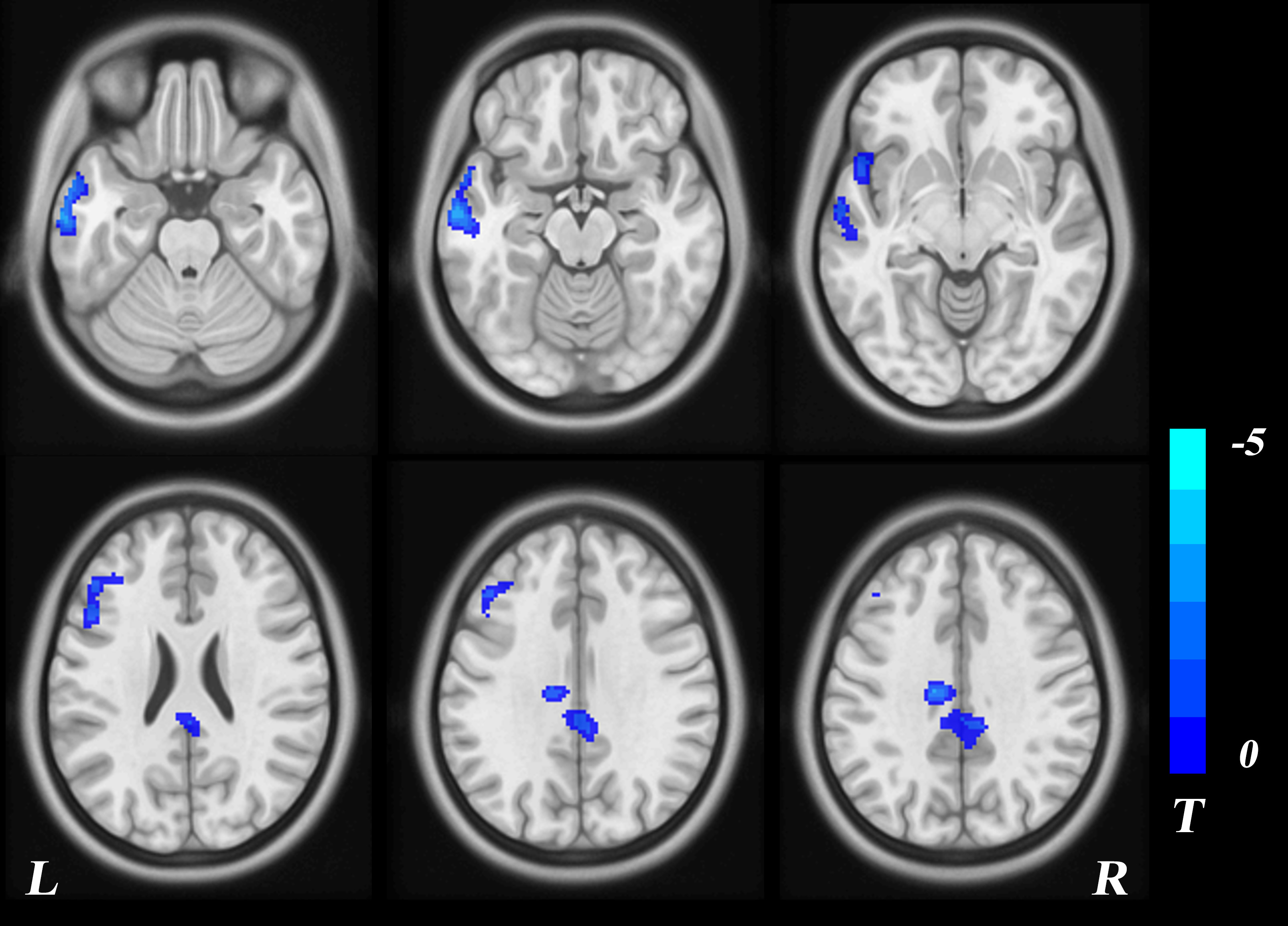
Figure 1. Regions showing CBF differences between T2DM patients and healthy controls (P<0.05, GRF corrected); the color-bar represented the T-value of the two-sample T-test, and the blue represented the lower CBF in T2DM patients than in HC group.
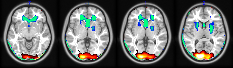
Figure 2. Regions showing cerebral perfusion networks discriminated T2DM patients and healthy controls (P<0.05). GIS1 (red-yellow) represented a perfusion characteristic network composed of bilateral superior occipital gyrus, cuneate lobe and superior parietal gyrus. GIS2 (blue-green) represented a perfusion characteristic network composed of bilateral anterior cingulate gyrus, caudate nucleus head, shell, et.al.