Nathan Davis1, Steven Baete2,3, Smita Rao4, Jill Slade5, Prodromos Parasoglou2,3, and Ryan Brown2,3
1New York Institute of Technology, Old Westbury, NY, United States, 2Department of Radiology, Bernard and Irene Schwartz Center for Biomedical Imaging, New York University Grossman School of Medicine, New York, NY, United States, 3Department of Radiology, Center for Advanced Imaging Innovation and Research, New York University Grossman School of Medicine, New York, NY, United States, 4Department of Physical Therapy, New York University, New York, NY, United States, 5Department of Radiology, Michigan State University, East Langsing, MI, United States
1New York Institute of Technology, Old Westbury, NY, United States, 2Department of Radiology, Bernard and Irene Schwartz Center for Biomedical Imaging, New York University Grossman School of Medicine, New York, NY, United States, 3Department of Radiology, Center for Advanced Imaging Innovation and Research, New York University Grossman School of Medicine, New York, NY, United States, 4Department of Physical Therapy, New York University, New York, NY, United States, 5Department of Radiology, Michigan State University, East Langsing, MI, United States
Diffusion tensor imaging can distinguish microstructural changes in the
tibial nerve of patients with diabetic peripheral neuropathy, but did not
detect longitudinal change in response to short-term exercise.
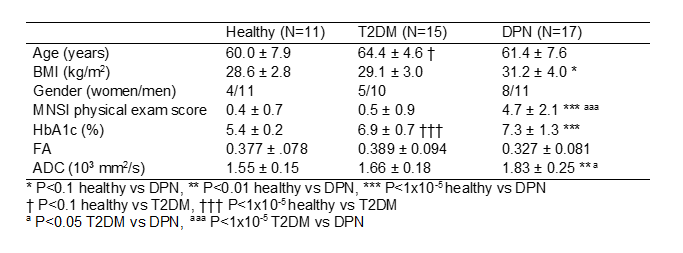
Table 1.
Participant
characteristics, diabetic markers, and DTI markers for the cross-sectional
study.
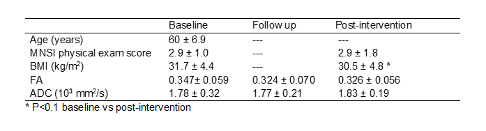
Table 2. Participant
characteristics, diabetic markers, and DTI markers for the longitudinal study (N=16).