Geon-Ho Jahng1,2, Chang Hyun Yoo3, Seokha Jin4, DongKyu Lee4, and HyungJoon Cho4
1Radiology, Kyung Hee University Hospital at Gangdong, Seoul, Korea, Republic of, 2Medicine, Kyung Hee University, Seoul, Korea, Republic of, 3Physics, Kyung Hee University, Seoul, Korea, Republic of, 4Biomedical Engineering, Ulsan National Institute of Science and Technology, Ulsan, Korea, Republic of
1Radiology, Kyung Hee University Hospital at Gangdong, Seoul, Korea, Republic of, 2Medicine, Kyung Hee University, Seoul, Korea, Republic of, 3Physics, Kyung Hee University, Seoul, Korea, Republic of, 4Biomedical Engineering, Ulsan National Institute of Science and Technology, Ulsan, Korea, Republic of
All MV
indices were sensitive enough to map accumulations
of amyloid plaques. The MV indices were sensitive to changes
in microbleed loads and
microvessel size. Therefore,
we recommend evaluating MV structure changes in the AD human brain using 3T MRI
with a Gadolinium (Gd)
contrast agent.

Table 1. List of parameters used in the simulation
with the condition that only the vascular structure exists in the voxel
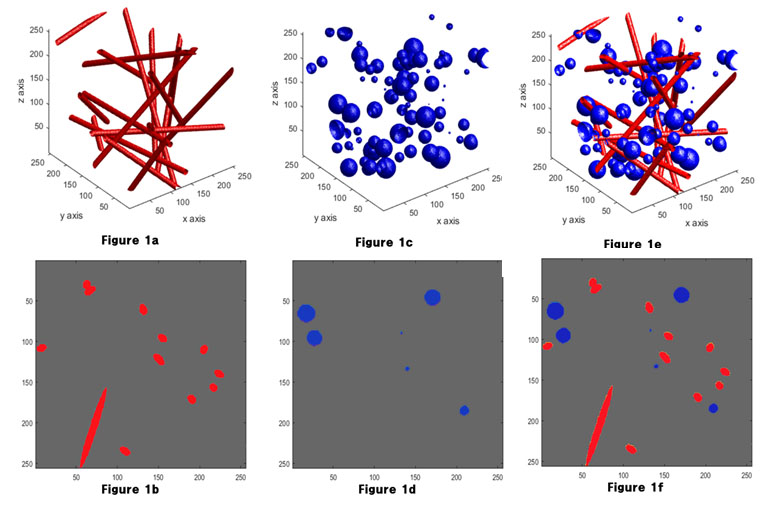
Figure 1. Simulation modeling
This figure shows the simulation models with 3D image of the modeled vascular structure (1a) and the corresponding cross-section taken along the z-axis (1b), 3D image of the modeled Aβ or microbleeds (1c) and the corresponding cross-section taken along the z-axis (1d), and 3D image of the modeled vascular structure with the amyloid-beta plaque or microbleed structure (1e) and the corresponding cross-section taken along the z-axis (1f).